Inflammation outside the brain may drive Parkinson’s disease. Researchers uncover a possible mechanism
A new study suggests that chronic inflammation may begin in peripheral tissues and then reach the brain through DNA-carrying extracellular vesicles, potentially contributing to neurodegeneration.
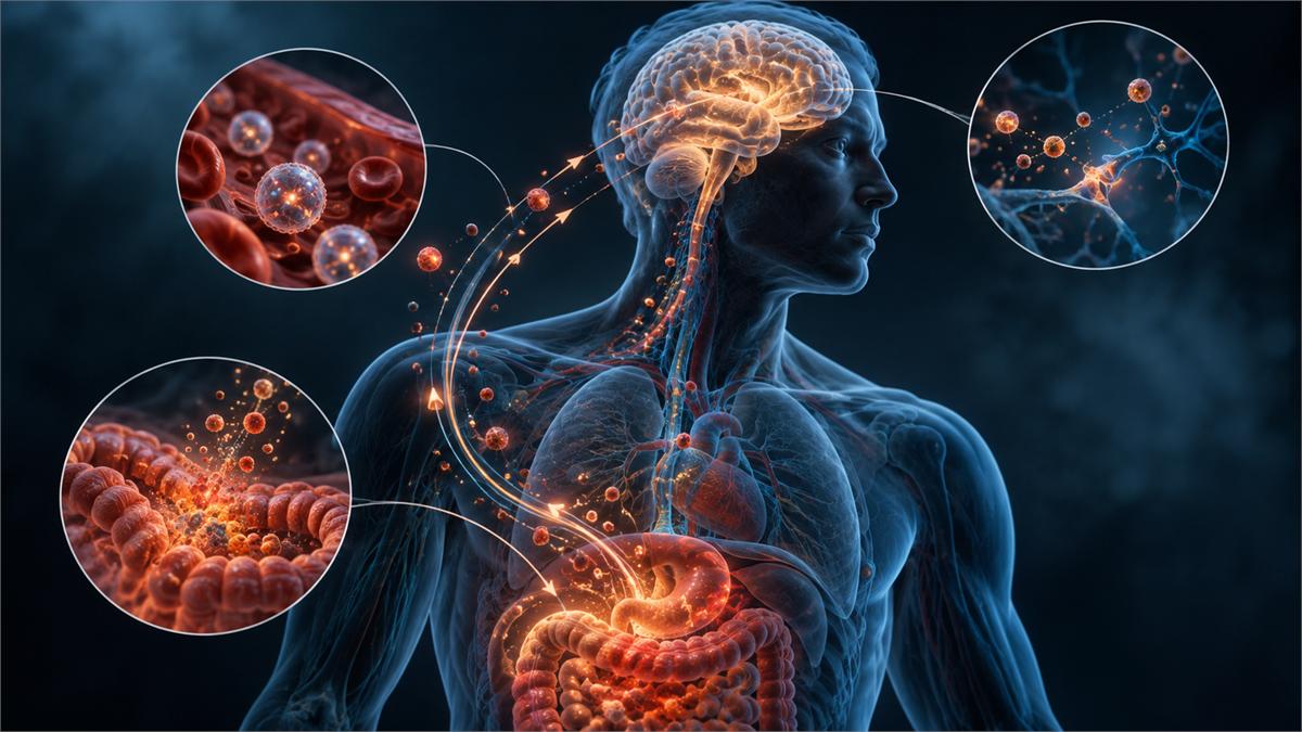
Table of contents
Inflammation may appear outside the brain first
Parkinson’s disease is primarily associated with the progressive loss of dopamine-producing neurons in the brain. A new study suggests, however, that part of the biological process contributing to neurodegeneration may begin earlier in peripheral tissues.
The researchers focused on chronic age-related inflammation, often referred to as inflammaging, and on the cGAS–STING immune pathway. This pathway helps cells detect DNA that appears in places where it normally should not be found.
With aging, or in the presence of certain variants of the LRRK2 gene, the cell’s own DNA may accumulate in the cytoplasm. According to the proposed mechanism, this can activate inflammatory signaling, while some of the DNA is then transported between cells inside small extracellular vesicles.
In the animal model, inflammatory changes in peripheral tissues appeared before similar changes in the brain and before deterioration in motor function. This temporal sequence is one of the main reasons the authors propose that inflammatory signaling may spread from the periphery toward the central nervous system.
Study details
The study combined observations from humans with experiments in animals and cells. This allowed the researchers to examine whether similar biological signals were present in people with Parkinson’s disease and to experimentally test parts of the proposed mechanism.
- Publication title: STING-dependent peripheral inflammaging drives neurodegeneration via extracellular vesicles.
- Authors: Maria Öberg, Caitlyn Myers, Najmeh Saffarzadeh and colleagues, from research teams including the University of Gothenburg and collaborating European institutions.
- Publication date: Published online on July 3, 2026, with the journal issue dated July 28, 2026.
- Journal: Cell Reports, volume 45, issue 7.
- DOI: 10.1016/j.celrep.2026.117640.
- PubMed: PMID 42397737.
- Study type: A mechanistic translational study combining human biological samples, an LRRK2 G2019S mouse model, cell experiments and genetic manipulation of the STING pathway.
- Population: The researchers analyzed samples from people with Parkinson’s disease as well as younger and older healthy controls. The material included plasma, cerebrospinal fluid and immune cells.
- Experimental model: Key causal experiments were performed in mice carrying the LRRK2 G2019S mutation, which increases LRRK2 activity and is associated with genetic risk of Parkinson’s disease.
- Processes analyzed: The researchers examined type I interferon signaling, cGAS–STING activity, DNA-containing extracellular vesicles, blood–brain barrier permeability, brain inflammation, motor function and the loss of dopaminergic neurons.
- Main mechanistic finding: The results suggest that aging-related dysfunction or excessive LRRK2 activity may lead to accumulation of the cell’s own DNA, its release inside extracellular vesicles and subsequent activation of the cGAS–STING pathway in other cells.
An important distinction is that human observations support the biological plausibility of the mechanism, while the strongest causal evidence comes from experiments in mice and cells.
How could inflammation reach the brain?
A central part of the proposed mechanism involves the system responsible for removing and recycling material inside cells. Its efficiency can decline with age. Similar dysfunction was observed with increased LRRK2 activity.
As a result, fragments of the cell’s own DNA may remain in the cytoplasm, where they are not normally expected to be present. The cell can interpret this misplaced DNA as a danger signal.
The sequence proposed by the researchers can be summarized as follows:
- Aging or increased LRRK2 activity impairs endolysosomal function, reducing the cell’s ability to degrade and recycle intracellular material.
- Nuclear and mitochondrial DNA begins to accumulate in the cytoplasm, where it can be detected by cGAS.
- The cGAS–STING pathway is activated, contributing to increased type I interferon signaling.
- Cells begin releasing more DNA-containing extracellular vesicles.
- These vesicles can reach other cells and activate STING-dependent immune responses again, allowing inflammatory signaling to spread through the body.
- In the mouse model, this process was accompanied by increased permeability of the blood–brain barrier, which may make it easier for peripheral inflammatory signals to affect the central nervous system.
Extracellular vesicles normally serve many useful communication functions. The problem may arise when they carry material capable of sustaining chronic immune activation.
Key findings
The study provided several independent lines of evidence supporting the same overall hypothesis. Its main strength lies in combining human observations with experiments that allowed researchers to manipulate individual components of the mechanism.
-
Inflammatory changes appeared first in peripheral tissues. In LRRK2 G2019S mice, a type I interferon signature was already detectable in peripheral tissues at 3 months of age, while comparable brain changes and worsening motor function appeared later, at around 12 months.
-
Removing STING reduced the effects associated with the mutation. Genetic deletion of STING normalized the elevated interferon response in examined peripheral cells and microglia and reduced signs of inflammation in the brain.
-
Blocking STING protected dopaminergic neurons in the animal model. Aging mice carrying the hyperactive LRRK2 variant lost more dopaminergic neurons than controls, whereas deletion of STING prevented this additional neuronal loss.
-
Extracellular vesicles carried biologically active DNA. Vesicles released by cells with increased LRRK2 activity contained more nuclear and mitochondrial DNA and were able to trigger STING-dependent signaling in recipient cells.
-
Similar signals were detected in people with Parkinson’s disease. Plasma and cerebrospinal fluid from patients contained more DNA-associated extracellular vesicles, and vesicles isolated from patients could activate STING-dependent interferon signaling in cell experiments.
Together, these results support a coherent biological model in which chronic inflammation may begin in peripheral tissues and then spread toward the brain through intercellular communication.
What could this mean for Parkinson’s disease?
Parkinson’s disease is not a single biological process and may arise through multiple pathways. LRRK2 mutations account for only a subset of cases, so these findings cannot automatically be generalized to every person with Parkinson’s disease.
The study is nevertheless relevant to the broader biology of aging. Endolysosomal dysfunction, accumulation of damaged DNA, chronic immune activation and increasing inflammaging are all processes that also occur during normal aging independently of LRRK2 mutations.
If the proposed mechanism is confirmed in future studies, it could point to several potential intervention targets:
- LRRK2 may represent a way to reduce the upstream processes that promote abnormal DNA accumulation and inflammatory signaling.
- The cGAS–STING pathway may offer another point at which chronic immune activation could potentially be interrupted.
- DNA-containing extracellular vesicles might eventually become biomarkers of disease-related inflammatory activity if their relevance is confirmed in larger clinical cohorts.
- Inflammation outside the central nervous system may prove to be a more important contributor to neurodegeneration than models focused mainly on processes occurring directly inside the brain have suggested.
This does not mean that there is currently a proven way to prevent Parkinson’s disease by blocking STING or reducing extracellular vesicles. The study identifies potential biological targets, not an established treatment.
Key limitations
The most informative parts of the study are the experiments that allowed researchers to manipulate individual components of the proposed pathway. At the same time, the experimental models limit how directly the findings can be applied to humans.
-
The strongest causal evidence comes from mice. Removing STING reduced neurodegeneration in the animal model, but it is not yet known whether targeting the same pathway would produce a similar effect safely in people with Parkinson’s disease.
-
The LRRK2 G2019S model represents only one part of Parkinson’s biology. LRRK2 is an important genetic risk factor, but most Parkinson’s disease cases are not caused by a single mutation in this gene.
-
Some experiments involving human-derived samples were based on relatively small sample sizes. These findings therefore require confirmation in larger and more diverse patient cohorts.
-
Detecting the same pathway in patients does not prove that it is the dominant cause of disease progression. Increased interferon signaling, endolysosomal dysfunction and DNA-containing extracellular vesicles could be drivers of disease, consequences of disease, or components of a broader network of interacting mechanisms.
For that reason, the results should not be interpreted as proof that Parkinson’s disease begins outside the brain in humans. Instead, the study provides a mechanistic model showing how such a process could occur and why it deserves further investigation.
Sources
Primary publication:
- Öberg M., Myers C., Saffarzadeh N. et al. STING-dependent peripheral inflammaging drives neurodegeneration via extracellular vesicles. Cell Reports. 2026;45(7):117640. DOI: 10.1016/j.celrep.2026.117640 · PubMed