Magnetic Resonance Imaging (3T MRI)
High-resolution imaging technique providing detailed views of soft tissues, brain, joints, and internal organs without radiation.
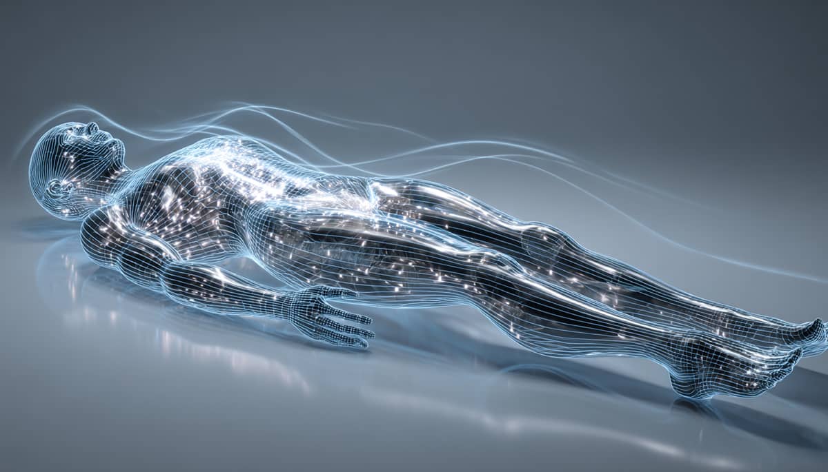
Table of contents
Basic data
Magnetic Resonance Imaging (MRI) is a non-invasive diagnostic technique that uses strong magnetic fields and radio waves to produce highly detailed images of the body’s internal structures. The 3 Tesla (3T) MRI offers significantly higher image resolution and faster scanning times compared to standard 1.5T devices.
MRI is a cornerstone of modern diagnostics — essential for identifying tissue damage, inflammation, structural abnormalities, and early-stage diseases in the brain, spine, joints, and internal organs. It provides unmatched contrast for soft tissue visualization without exposing the patient to ionizing radiation.
The 3T MRI is especially valuable for neurological, musculoskeletal, and cardiovascular imaging — supporting both preventive diagnostics and precise treatment planning.
Category: Imaging
Level: Beginner
Usefulness: High
Level
Beginner
Usefulness
High
High-resolution visualization of soft tissues
MRI offers unparalleled contrast and detail for soft tissues, helping detect injuries, inflammation, and early degenerative changes that X-rays or CT may miss.
Broad diagnostic range
Suitable for brain, spine, joint, cardiovascular, and organ imaging — allowing a single modality to cover multiple systems comprehensively.
Radiation-free safety
Unlike CT or X-ray, MRI uses no ionizing radiation, making it safe for repeated use and preventive applications.
How it works
Magnetic alignment
The MRI magnet aligns hydrogen nuclei (protons) within body tissues using a strong magnetic field.
Radiofrequency excitation
Short bursts of radio waves disturb this alignment, and as atoms realign, they emit measurable energy.
Image reconstruction
Advanced software converts these energy signals into detailed cross-sectional images, allowing visualization of soft tissue, organs, and structures.
Measures
Tissue structure and density
Visualizes muscles, ligaments, nerves, cartilage, and brain tissue in fine detail.
Inflammation and edema
Detects areas of fluid accumulation or tissue swelling associated with injury or disease.
Lesions and structural abnormalities
Identifies cysts, tumors, herniations, or degenerative changes at early stages.
Reliability
High diagnostic accuracy
3T MRI provides exceptional image clarity, reducing false negatives and enhancing diagnostic precision.
Operator and interpretation dependent
Reliability depends on proper calibration, patient stillness, and expert radiological interpretation.
Limitations
Cost and accessibility
MRI scans are more expensive and less widely available than X-rays or ultrasound.
Motion artifacts
Even small movements can blur the image, requiring patient cooperation and sometimes sedation for children.
Contraindications
Not suitable for individuals with pacemakers, certain metal implants, or severe claustrophobia.
Frequency
Suggested cadence
MRI should be performed when clinically indicated — typically every 1–3 years for monitoring known conditions or when new symptoms arise.
Cost
Typical costs
Prices vary by region and body part scanned. Typical range: $300–$800 for standard MRI, and $800–$1,200 for advanced 3T imaging with contrast.
Availability
Where available
Available in hospitals, imaging centers, and diagnostic clinics equipped with 1.5T or 3T MRI systems. 3T units are typically found in advanced diagnostic or research facilities.
Preparation
How to prepare
Avoid metal accessories and inform staff of any implants. Fasting may be required for abdominal scans. Remain still during imaging to ensure clear results.
Interpretation
Normal vs. abnormal findings
Reports describe normal anatomy and note deviations such as lesions, inflammation, or degeneration.
Comparative interpretation
Comparing multiple scans over time helps assess disease progression or recovery.
Alternatives
Computed Tomography (CT)
Faster and cheaper alternative for bone and lung imaging, but uses ionizing radiation and has lower soft tissue contrast.
Ultrasound
Safe and inexpensive for superficial organs but limited by depth and image resolution.
FAQ
What makes 3T MRI different from standard MRI?
The 3T scanner has twice the magnetic strength of a 1.5T system, providing higher-resolution images and shorter scan times — particularly useful for brain and joint imaging.
Is MRI safe?
Yes, MRI is safe for most people. It does not use radiation, but metal implants or devices may require special precautions.