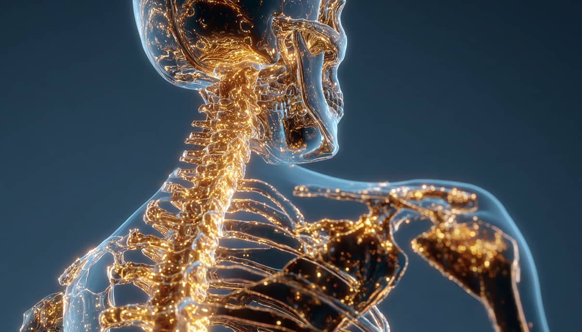
Gęstość mineralna kości (BMD)
Gęstość mineralna kości (BMD) — kluczowy biomarker siły szkieletu i zdrowego starzenia się
Spis treści
Przegląd
Gęstość mineralna kości (BMD) mierzy zawartość minerałów w jednostce powierzchni kości, najczęściej ocenianą metodą dwuenergetycznej absorpcjometrii rentgenowskiej (DXA) w odcinku lędźwiowym kręgosłupa, biodrze lub całym ciele. W przeciwieństwie do wskaźników T-score i Z-score — które pokazują, jak wynik jednostki wypada w porównaniu z młodymi zdrowymi dorosłymi (T) lub rówieśnikami w tym samym wieku (Z) — BMD jest surową wartością raportowaną przez urządzenie w g/cm², używaną do klasyfikacji osteopenii i osteoporozy oraz do monitorowania zmian w czasie.
Fizjologicznie BMD odzwierciedla wynik netto procesu przebudowy kości (tworzenie osteoblastów versus resorpcja osteoklastów), który jest regulowany przez obciążenia mechaniczne, hormony (np. estrogen, testosteron, PTH), odżywianie (wapń, witamina D, białko) oraz czynniki ogólnoustrojowe, takie jak stany zapalne czy leki. Wraz z wiekiem — szczególnie po menopauzie — resorpcja kości zaczyna przeważać nad ich tworzeniem, co prowadzi do obniżenia BMD i zwiększenia ryzyka złamań. Oprócz złamań, niższe BMD często współwystępuje z mniejszą masą i siłą mięśniową, niższą aktywnością fizyczną oraz podwyższonym ryzykiem kardiometabolicznym, co czyni je istotnym biomarkerem zarówno integralności szkieletu, jak i ogólnej długości zdrowia.
Kategoria: Kości
Wpływ: Wysoki
Zakresy referencyjne
Uwaga:Prezentowane zakresy referencyjne opierają się na danych populacyjnych. Wartości mogą się różnić w zależności od badania lub zbioru danych. Przedstawione zakresy to uśrednienia z wielu źródeł naukowych. Pełna lista źródeł znajduje się w sekcji Dane naukowe.
Zakresy referencyjne populacji — Mężczyźni (Gęstość mineralna kości, g/cm²)
| Percentyl | Przedziały wiekowe | |||||
|---|---|---|---|---|---|---|
| 20–29 | 30–39 | 40–49 | 50–59 | 60–69 | 70–79 | |
| 90 (Doskonały) | 1.45 | 1.45 | 1.4 | 1.3 | 1.2 | 1.1 |
| 80 | 1.4 | 1.4 | 1.35 | 1.25 | 1.15 | 1.05 |
| 70 (Powyżej średniej) | 1.35 | 1.35 | 1.3 | 1.2 | 1.1 | 1 |
| 60 | 1.3 | 1.3 | 1.25 | 1.15 | 1.05 | 0.95 |
| 50 (Średni) | 1.25 | 1.25 | 1.2 | 1.1 | 1 | 0.9 |
| 40 | 1.2 | 1.2 | 1.15 | 1.05 | 0.95 | 0.85 |
| 30 (Poniżej średniej) | 1.15 | 1.15 | 1.1 | 1 | 0.9 | 0.8 |
| 20 | 1.1 | 1.1 | 1.05 | 0.95 | 0.85 | 0.75 |
| 10 (Słaby) | 1.05 | 1.05 | 1 | 0.9 | 0.8 | 0.7 |
Zakresy referencyjne populacji — Kobiety (Gęstość mineralna kości, g/cm²)
| Percentyl | Przedziały wiekowe | |||||
|---|---|---|---|---|---|---|
| 20–29 | 30–39 | 40–49 | 50–59 | 60–69 | 70–79 | |
| 90 (Doskonały) | 1.22 | 1.2 | 1 | 0.98 | 0.94 | 0.92 |
| 80 | 1.2 | 1.18 | 0.98 | 0.95 | 0.9 | 0.88 |
| 70 (Powyżej średniej) | 1.18 | 1.15 | 0.96 | 0.93 | 0.88 | 0.85 |
| 60 | 1.16 | 1.12 | 0.94 | 0.9 | 0.86 | 0.82 |
| 50 (Średni) | 1.14 | 1.1 | 0.92 | 0.88 | 0.84 | 0.8 |
| 40 | 1.12 | 1.08 | 0.9 | 0.86 | 0.82 | 0.78 |
| 30 (Poniżej średniej) | 1.1 | 1.06 | 0.88 | 0.84 | 0.8 | 0.76 |
| 20 | 1.08 | 1.04 | 0.86 | 0.82 | 0.78 | 0.74 |
| 10 (Słaby) | 1.06 | 1.02 | 0.84 | 0.8 | 0.76 | 0.72 |
Populacja w zdrowym zakresie
Ogólnie rzecz biorąc, duża część dorosłej populacji utrzymuje wartości gęstości mineralnej kości w zakresie średnim lub powyżej średniej do połowy życia, po czym stopniowo zaczyna się ich spadek. Różnice między mężczyznami a kobietami pojawiają się głównie w wyniku zmian hormonalnych i fizjologicznych, które wpływają na przebudowę kości.
Młodsi dorośli
Większość osób w wieku dwudziestu i trzydziestu lat utrzymuje wartości BMD w zakresie od średniego do doskonałego, co odzwierciedla osiągnięcie szczytowej masy kostnej.
Kobiety po menopauzie
Po menopauzie obserwuje się gwałtowny spadek BMD w wyniku obniżenia poziomu estrogenu. Mniejszy odsetek kobiet utrzymuje zdrowe wartości bez interwencji stylu życia lub leczenia.
Starsze osoby dorosłe
U obu płci po 60. roku życia występuje postępujący spadek BMD, choć mężczyźni zazwyczaj zachowują wyższe wartości bezwzględne niż kobiety.
| Grupa populacyjna | Zdefiniowany optymalny zakres | Odsetek w zakresie |
|---|---|---|
| Młodzi dorośli (20–39 lat) | ≥1,10 g/cm² (kręgosłup lędźwiowy lub całe ciało) | Większość w zakresie średnim lub powyżej średniej |
| Dorośli w średnim wieku (40–59 lat) | ≥1,00 g/cm² | Umiarkowany odsetek w zdrowym zakresie |
| Starsze osoby dorosłe (60+) | ≥0,85 g/cm² | Mniejszy odsetek utrzymuje optymalną gęstość kości |
Ogólnie dane populacyjne wskazują, że choć wiele osób osiąga i utrzymuje zdrowe wartości BMD w młodym wieku dorosłym, gęstość kości zwykle spada wraz z wiekiem, szczególnie u kobiet po menopauzie. Utrzymanie optymalnego zdrowia kości wymaga aktywności fizycznej, odpowiedniego odżywiania i regularnych badań profilaktycznych przez całe życie.
Wpływ na zdrowie i długowieczność
Wpływ: Wysoki
Kluczowy wniosek
Gęstość mineralna kości (BMD) stanowi potężny biomarker zdrowia ogólnoustrojowego i biologicznego starzenia. Niska BMD przewiduje nie tylko ryzyko złamań, ale także wyższą śmiertelność całkowitą, choroby sercowo-naczyniowe i pogorszenie funkcji poznawczych, podczas gdy wyższa BMD wiąże się z dłuższym życiem i mniejszym obciążeniem chorobami przewlekłymi. Badania populacyjne na różnych kontynentach konsekwentnie pokazują, że utrzymanie optymalnej BMD wspiera samodzielność funkcjonalną i wydłużoną długość zdrowia.
Związek z długowiecznością
Wyższa BMD jest silnie i niezależnie powiązana z dłuższą długością życia i niższym ryzykiem śmierci. Duże badania kohortowe w USA, na Tajwanie i w Europie pokazują, że osoby z niskim BMD mają o 30–70% wyższe ryzyko przedwczesnej śmierci, nawet po uwzględnieniu czynników zakłócających, takich jak wiek, BMI czy choroby współistniejące. Z kolei utrzymywanie BMD w wyższych percentylach wiąże się ze zwiększonym przeżyciem w różnych populacjach i płciach.
Mechanizmy
Związek między BMD a długowiecznością jest pośredniczony przez kilka szlaków biologicznych: zmniejszoną śmiertelność związaną ze złamaniami, niższy poziom stanu zapalnego, lepszy profil sercowo-naczyniowy i metaboliczny, a także wspólne czynniki, takie jak siła mięśni, mobilność i równowaga hormonalna. Tkanka kostna działa również jako narząd endokrynny, wpływając na metabolizm energetyczny i zdrowie naczyń.
Podsumowanie praktyczne
Utrzymywanie gęstości kości poprzez trening oporowy, odpowiednią podaż wapnia i witaminy D, optymalne spożycie białka oraz unikanie palenia i nadmiernego spożycia alkoholu znacząco poprawia długość zdrowia. Regularne badania BMD, szczególnie po 40. roku życia lub po menopauzie, umożliwiają wczesną interwencję w celu zapobiegania spadkom i utrzymania długoterminowej odporności szkieletowej i ogólnoustrojowej.
Jak często mierzyć
BMD należy okresowo mierzyć, aby monitorować zdrowie kości i wykrywać zmiany w czasie. Częstotliwość zależy od poziomu ryzyka, wieku oraz prowadzonych interwencji.
Populacja ogólna
Co 3–5 lat, począwszy od około 40–50 roku życia, lub wcześniej, jeśli występują czynniki ryzyka (np. wywiad rodzinny, niski BMI, menopauza).
Osoby z grupy ryzyka lub w trakcie leczenia
Co 1–2 lata w celu oceny skuteczności terapii lub zmian gęstości kości po interwencji.
Sportowcy i osoby aktywne fizycznie
Co 2–3 lata w celu monitorowania wpływu treningu i odżywiania na zdrowie kości.
Strategie poprawy
Trening oporowy i obciążeniowy
Regularnie wykonuj ćwiczenia siłowe i z obciążeniem, aby stymulować tworzenie kości i spowalniać ich resorpcję.
Optymalizacja spożycia wapnia, witaminy D i białka
Zadbaj o odpowiednią podaż tych składników z diety lub suplementów, aby wspierać przebudowę i mineralizację kości.
Wsparcie równowagi hormonalnej i metabolicznej
Utrzymuj prawidłową masę ciała, odpowiednią ilość snu i kontroluj stres; rozważ konsultację medyczną w kierunku niedoborów hormonalnych.
FAQ
Jak BMD wpływa na długowieczność i ogólny stan zdrowia?
Niska BMD wiąże się nie tylko z ryzykiem złamań, ale także z wyższą śmiertelnością i obciążeniem chorobami przewlekłymi, co czyni ją kluczowym biomarkerem długowieczności.
Dlaczego BMD spada z wiekiem?
Połowa życia to moment, w którym resorpcja kości zaczyna przyspieszać — zwłaszcza po menopauzie — z powodu zmian hormonalnych, mniejszej aktywności fizycznej i niedostatecznej podaży składników odżywczych.
Czy można naturalnie poprawić BMD?
Tak. Trening oporowy, optymalne odżywianie, wystarczająca ilość witaminy D oraz styl życia ograniczający stan zapalny pomagają poprawić lub utrzymać gęstość kości.
Czy wysoka BMD zawsze jest korzystna?
Zazwyczaj tak, jeśli chodzi o wytrzymałość kości, jednak ekstremalnie wysokie wartości mogą czasami wiązać się z ryzykiem metabolicznym; najlepiej utrzymywać poziom w optymalnym zakresie.
Dane naukowe
The associations between bone mineral density and long-term risks of cardiovascular disease, cancer, and all-cause mortality
Typ badania: non-rct observational study
Liczba cytowań: 18
Rok: 2022
Autorzy: Lin Shi, Xiao Yu, Q. Pang, Xian-Jun Chen, Cheng-Hao Wang
Czasopismo: Frontiers in Endocrinology
Ranking czasopisma: Q1
Główne wnioski: Osteoporosis is associated with an increased risk of all-cause mortality, particularly in older individuals and those with lower BMI.
Streszczenie: Objective We aimed to investigate the associations between bone mineral density and long-term risks of cardiovascular disease (CVD), cancer, and all-cause mortality in nationwide survey participants aged 18 and over. Methods Using data from the United States National Health and Nutrition Examination Survey III (NHANES III), the associations of bone mineral density (normal bone mass, osteopenia, and osteoporosis) with CVD, cancer, and all-cause mortality were analyzed using the Cox proportional hazards model. Results A total of 11,909 adults aged 18 and over were enrolled in this study. Compared with the participants with normal bone mass, those with osteoporosis and osteopenia were more likely to be female, of non-Hispanic white ethnicity, and older. They were also more likely to have lower calcium and vitamin D intakes, a lower body mass index (BMI), lower educational attainment, and lower family incomes. Participants with osteoporosis and osteopenia also engaged in less physical activity and were more likely to have diabetes, high blood pressure, and a history of CVD. After adjusting for confounders, osteopenia and osteoporosis were significantly associated with all-cause mortality, with the hazard ratios (95% confidence intervals) being 1.37 (1.11, 1.68) and 1.06 (0.91, 1.25), respectively, compared with normal bone mass. Age (P for interaction = 0.001) and BMI (P for interaction = 0.002) were found to modify the association between bone mineral density and all-cause mortality. Conclusions In a nationally representative cohort, osteoporosis was associated with an increased risk of all-cause mortality, and this association was stronger in participants who were older and had a lower BMI.
Zobacz badanieAssociation between bone mineral density and cardiovascular disease in older adults
Typ badania: non-rct observational study
Liczba cytowań: 19
Rok: 2023
Autorzy: Yulu Yang, Yun Huang
Czasopismo: Frontiers in Public Health
Ranking czasopisma: Q1
Główne wnioski: Higher femur bone mineral density is associated with a lower risk of cardiovascular disease in older adults over 60 years old, with an inflection point of 0.741 gm/cm2.
Streszczenie: Background and aims Cardiovascular disease and osteoporosis are common diseases in older adults with high morbidity. The study on the interaction between the two in pathogenic mechanisms has been paid much attention by the majority of researchers. This study aimed to explore the relationship between bone mineral density and cardiovascular disease in older adults. Methods The primary data was downloaded from the National Health and Nutrition Examination Survey database of the United States. Multivariate logistic regression model, generalized additive model, and smooth curve fitting were used to explore the relationship between bone mineral density and cardiovascular events risk. When a curve relationship was found, a two-piecewise linear model was used to calculate the inflection point. In addition, subgroup analysis was also performed. Results A total of 2097 subjects were included in this study. After adjusting for potential confounders, no significant association was found between lumbar bone mineral density and cardiovascular disease, while femur bone mineral density had a non-linear relationship with cardiovascular disease, with an inflection point of 0.741 gm/cm2. When bone mineral density was <0.741 gm/cm2, the risk of cardiovascular disease decreased speedily. Once bone mineral density exceeded this value, the risk of cardiovascular disease continued to decrease, but the trend became significantly slower. Compared with patients with normal bone mass, osteoporosis was associated with a 2.05-fold increased risk of cardiovascular disease (95% CI 1.68–5.52). There were no significant differences in interaction tests of all subgroups (p for interaction >0.05) except race. Conclusion Our results indicated that bone mineral density was closely associated with the prevalence of cardiovascular disease in older adults over 60 years old, especially the femur bone mineral density was negatively non-linear associated with cardiovascular disease risk, with an inflection point of 0.741 gm/cm2.
Zobacz badanieGenetic predisposition to bone mineral density and their health conditions in East Asians.
Typ badania: non-rct observational study
Liczba cytowań: 6
Rok: 2024
Autorzy: Ying-Ju Lin, Wen-Miin Liang, Jian-Shiun Chiou, Chen-Hsing Chou, Ting-Yuan Liu, Jai-Sing Yang, Te-Mao Li, Y. Fong, I. Chou, Ting-Hsu Lin, Chiu‐Chu Liao, Shao-Mei Huang, Fuu-Jen Tsai
Czasopismo: Journal of bone and mineral research : the official journal of the American Society for Bone and Mineral Research
Ranking czasopisma: Q1
Główne wnioski: Genetic factors influence bone mineral density in East Asians, with higher BMI increasing BMD but no direct causal relationship found between BMD and type 2 diabetes or osteoporosis.
Streszczenie: Osteoporosis, a condition defined by low bone mineral density (BMD) (typically < -2.5 SD), cause a higher fracture risk and lead to significant economic, social, and clinical impacts. Genome-wide studies mainly in Caucasians have found many genetic links to osteoporosis, fractures, and BMD, with limited research in East Asians. We investigated the genetic aspects of BMD in 86,716 individuals from the Taiwan Biobank and their causal links to health conditions within East Asians. A genome-wide association study (GWAS) was conducted, followed by observational studies, polygenic risk score assessments, and genetic correlation analyses to identify associated health conditions linked to BMD. GWAS and gene-based GWAS studies identified 78 significant SNPs and 75 genes related to BMD, highlighting pathways like Hedgehog, WNT-mediated, and TGF-β. Our cross-trait linkage disequilibrium score regression analyses for BMD and osteoporosis consistently validated their genetic correlations with body mass index (BMI) and type 2 diabetes (T2D) in East Asians. Higher BMD was linked to lower osteoporosis risk but increased BMI and T2D, whereas osteoporosis linked to lower BMI, waist circumference, HbA1c, and reduced T2D risk. Bidirectional Mendelian randomization (MR) analyses revealed that a higher BMI causally increases BMD in East Asians. However, no direct causal relationships were found between BMD and T2D, or between osteoporosis and either BMI or T2D. This study identified key genetic factors for bone health in Taiwan, and revealed significant health conditions in East Asians, particularly highlighting the genetic interplay between bone health and metabolic traits like T2D and BMI.
Zobacz badanieAssociation of low bone mineral density and dementia in older women: insights from the Longevity Improvement and Fair Evidence Study.
Typ badania:
Liczba cytowań: 1
Rok: 2025
Autorzy: K. Kawaguchi, M. Maeda, Fumiko Murata, Yasuharu Nakashima, Haruhisa Fukuda
Czasopismo: Age and ageing
Ranking czasopisma: Q1
Główne wnioski: Low bone mineral density is associated with a higher risk of developing dementia in older women aged 65 years, suggesting that osteoporosis screenings could be useful for both secondary and primary prevention of dementia.
Streszczenie: BACKGROUND Both osteoporosis and dementia have emerged as important public health challenges in Japan's aging population. This study aimed to investigate the impact of low bone mineral density (BMD) on the subsequent risk of dementia in older Japanese women aged ≥65 years, given the overlapping demographics of individuals affected by these two conditions. METHODS This cohort study was conducted using osteoporosis screening data and insurance claims data from a municipality. We identified 8618 women (median age: 73 years) who underwent osteoporosis screening between April 2019 and March 2021. Participants with a BMD <80% of the young adult mean were assigned to a low-BMD group (n = 2297), whereas those with a BMD ≥80% were assigned to a control group (n = 6321). The study outcomes were new-onset all-cause dementia and Alzheimer's disease (AD). To estimate the risk of low BMD on these outcomes, we constructed Cox proportional hazards models that adjusted for covariates (age, care needs, year of cohort entry, comorbidities and medications) using inverse probability of treatment weighting. RESULTS The low-BMD group had a significantly higher risk of developing both all-cause dementia (adjusted hazard ratio: 1.58, 95% confidence interval: 1.20-2.08) and AD (1.61, 1.11-2.36) than the control group over approximately 30 months of follow-up. CONCLUSION These findings suggest that low BMD is associated with medium-term onset of dementia. Osteoporosis screenings could be useful not only for the secondary prevention of osteoporosis, but also for the primary prevention of dementia.
Zobacz badanieAssociation between life’s essential 8 and bone mineral density among adults aged 20–59 years
Typ badania: non-rct observational study
Liczba cytowań: 1
Rok: 2025
Autorzy: Yuyu Cui, Zhening Xu, Zhaoshu Cui, Yuanyuan Guo, Peiwei Wu, Xiaoyan Zhou
Czasopismo: Scientific Reports
Ranking czasopisma: Q1
Główne wnioski: Higher Life's Essential 8 scores and cardiovascular health are linked to greater bone mineral density in adults aged 20-59 years.
Zobacz badanieDietary Patterns in Relation to Low Bone Mineral Density and Fracture Risk: A Systematic Review and Meta-Analysis.
Typ badania: meta-analysis
Liczba cytowań: 113
Rok: 2019
Autorzy: R. Fabiani, Giulia Naldini, M. Chiavarini
Czasopismo: Advances in nutrition
Ranking czasopisma: Q1
Główne wnioski: The 'Healthy' and 'Milk/dairy' dietary patterns are associated with a reduced risk of low bone mineral density and fractures, while the 'Western' pattern increases these risks.
Streszczenie: Low bone mineral density (BMD) and osteoporosis-related fractures constitute a considerable public health burden. Several studies have demonstrated the association between diet and bone health. We performed a systematic review to provide an estimate of the association between different dietary patterns defined through the use of a posteriori methods and fracture or low BMD risk. A literature search on PubMed, Web of Science, and Scopus databases, up to March 2018, was performed to identify all eligible case-control, prospective, or cross-sectional studies involving subjects of both sexes and any age. Random-effects models were used. Heterogeneity and publication bias were evaluated. Stratified analyses were conducted on study characteristics. The meta-analysis includes 20 studies and identifies 3 prevalent dietary patterns: 'Healthy,' 'Milk/dairy,' and 'Meat/Western.' From the 10 studies on fracture, adherence to the 'Healthy' pattern reduced the risk, particularly in older people (OR: 0.79; 95% CI: 0.66, 0.95; P = 0.011) and in Eastern countries (OR: 0.64; 95% CI: 0.43, 0.97; P = 0.037), whereas the risk increased with the 'Meat/Western' pattern, especially for older people (OR: 1.11; 95% CI: 1.04, 1.18, P = 0.001), in those with hip fractures (OR: 1.15; 95% CI: 1.05, 1.25; P = 0.002), and in Western countries (OR: 1.10; 95% CI: 1.07, 1.14; P < 0.0001). Analyses on low BMD showed a reduced risk in the 'Healthy' pattern, particularly for younger people (OR: 0.62; 95% CI: 0.44, 0.89; P = 0.009). The 'Meat/Western' pattern increased low BMD risk, especially in older people (OR: 1.31; 95% CI: 1.05, 1.64; P = 0.015). The 'Milk/dairy' pattern resulted in the strongest reduction in low BMD risk; when stratifying, this effect remained significant (e.g., older women-OR: 0.57; 95% CI: 0.46, 0.70; P < 0.0001). Nutrition is an important modifiable factor affecting bone health. The 'Healthy' and 'Milk/dairy' patterns are associated with a reduced risk of low BMD and fracture. In contrast, the 'Western' pattern is inversely associated.
Zobacz badanieRelationship between bone mineral density and oral health: a cross sectional observational study
Typ badania: non-rct observational study
Liczba cytowań: 0
Rok: 2025
Autorzy: Rahime Zeynep Erdem, Mustafa Erdem, Mustafa Kıranatlı, Kevser Karakaya
Czasopismo: BMC Oral Health
Ranking czasopisma: Q1
Główne wnioski: Tooth loss and dental decay rates are significantly higher in patients with osteoporosis, and bone resorption during osteopenia is a crucial risk factor for dental health.
Streszczenie: Purpose Bone mineral density (BMD) is related to oral health. This study investigated how changes in BMD influence tooth loss risk and dental caries prevalence. Methods This cross-sectional observational study included 224 people (199 males and 25 females). The BMD scores of the participants’ lumbar spine, femoral neck, and total hip were categorized as normal, osteopenia, or osteoporosis. Oral health was assessed using the Decayed, Missing, Filled Teeth (DMFT) index and Oral Hygiene Index-Simplified (OHI-S) scores. Based on the number of surviving teeth, the participants were categorized into low (< 20) and high (≥ 20) groups. Differences between groups were assessed using independent sample T tests and one-way analysis of variance. Results The normal, osteopenia, and osteoporosis groups comprised 72, 87, and 65 participants, respectively. The OHI-S scores showed no notable variations across the groups. The DMFT index scores were highest (18.69) in the osteoporosis group and lowest (14.08) in the normal group (p < 0.001). Although the number of remaining teeth was lower in the osteoporosis and osteopenia groups compared to the normal group (p < 0.001), that in the osteopenia group approximated the normal group, but was substantially higher than in the osteoporosis group. The group with the lowest number of remaining teeth had lower total hip T-scores, despite significantly higher DMFT indexes (p < 0.001). Conclusions Tooth loss and dental decay rates were significantly high in patients with osteoporosis. Although bone resorption during osteopenia is not excessive, it constitutes a crucial risk factor for dental health. Therefore, attention must be paid to bone resorption treatment in patients with osteopenia.
Zobacz badanieBone mineral density and the risk of kidney disease in patients with type 1 diabetes.
Typ badania: non-rct observational study
Liczba cytowań: 2
Rok: 2024
Autorzy: Sabina Chaudhary Hauge, H. Ø. Hjortkjær, Frederik Persson, S. Theilade, Morten Frost, N. R. Jørgensen, P. Rossing, Ditte Hansen
Czasopismo: Journal of diabetes and its complications
Ranking czasopisma: Q1
Główne wnioski: Low bone mineral density is associated with the progression of diabetic kidney disease in patients with type 1 diabetes mellitus, suggesting an interaction between bone and kidney.
Zobacz badanieRisk factors and renal outcomes of low bone mineral density in patients with non-dialysis chronic kidney disease
Typ badania: non-rct observational study
Liczba cytowań: 12
Rok: 2020
Autorzy: Y. Hyun, K.-B. Lee, S. Han, K. H. Choi, H. Park, Y. Oh, S. Park, K. Oh, C. Ahn, on behalf of the KoreaN cohort study for Outcome in p Group
Czasopismo: Osteoporosis International
Ranking czasopisma: Q1
Główne wnioski: Low bone mineral density is associated with poor renal outcomes in non-dialysis chronic kidney disease, with modifiable lifestyle factors like low physical activity and high dietary Na/K intake ratio playing a role.
Streszczenie: Summary Bone disorder is a common complication of chronic kidney disease (CKD). The clinical usefulness of bone mineral density (BMD) in CKD is not well known. Our study shows that low BMD is associated with physical activity and dietary Na/K intake ratio and can predict poor renal outcome in non-dialysis CKD. Purpose Despite evidence of a link between bone mineral disorders and chronic kidney disease (CKD), the clinical implications of bone mineral density (BMD) in CKD are not well established. We investigated risk factors and renal outcomes of low BMD in CKD. Methods We analyzed data from the KNOW-CKD. BMD measured by dual-energy x-ray absorptiometry was classified by T score: normal ( T score ≥ − 1.0), osteopenia (− 1.0 > T score > − 2.5), and osteoporosis ( T score ≤ − 2.5) of the lumbar spine, hip, or femoral neck. Logistic regression analysis to assess risk factors of low BMD ( T score < − 1.0) and Cox proportional hazards models to estimate risk of incident end-stage renal disease (ESRD). Results Low BMD was prevalent (osteopenia 33%; osteoporosis 8%) in 2128 adults with CKD (age 54 ± 12 years; male 61%). Over a median follow-up of 4.3 years, there were 521 cases of incident ESRD. Lower BMD was associated with female sex, older age, low eGFR, low BMI, and lifestyle factors of physical activity (odds ratio (OR) = 0.62, 95% confidence interval (0.49–0.77)) and spot urine Na/K ratio (1.07 (1.00–1.15)). In adjusted Cox models, low BMD was associated with increased incident ESRD (hazard ratio (HR) = 1.14 (0.92–1.41) for osteopenia; 1.43 (1.01–2.04) for osteoporosis, P for trend < 0.05) compared with the reference of normal BMD. The association between low BMD and ESRD was similar according to T score discordance classification. Conclusions Low BMD was associated with modifiable lifestyle factors including low physical activity and high dietary Na/K intake ratio. The presence of low BMD is associated with poor renal outcomes in non-dialysis CKD.
Zobacz badanieBone mineral density and osteoporosis in relation to all-cause and cause-specific mortality in NHANES: a population-based cohort study.
Typ badania:
Liczba cytowań: 47
Rok: 2020
Autorzy: S. Cai, Jiayao Fan, Lina Zhu, Jianhong Ye, Xianming Rao, C. Fan, Y. Zhong, Yingjun Li
Czasopismo: Bone
Ranking czasopisma: Q1
Główne wnioski: Maintaining normal bone mineral density is crucial to lower the risk of mortality, with higher BMD levels in femur being associated with decreased risk of cancer and heart diseases mortality.
Zobacz badanieThe association between bone mineral density and risk of mortality: A prospective cohort study of 233,397 Taiwanese.
Typ badania: non-rct observational study
Liczba cytowań: 0
Rok: 2024
Autorzy: Honglin Cai, Tsung Yu, Timothy Kwok, Samuel Yeung Shan Wong, Martin C S Wong, Xiang Qian Lao
Czasopismo: Bone
Ranking czasopisma: Q1
Główne wnioski: Low bone mineral density (BMD) is associated with an increased risk of all-cause, cardiovascular disease, and cancer mortality in both men and women, with a stronger positive association in women.
Zobacz badanieTen-year atherosclerotic cardiovascular disease risk score in post-menopausal women with low bone mineral density
Typ badania: non-rct observational study
Liczba cytowań: 1
Rok: 2025
Autorzy: K. Wani, S. Sabico, Nicola Veronese, Abeer A. Al-Masri, N. Al-Daghri
Czasopismo: Aging Clinical and Experimental Research
Ranking czasopisma: Q2
Główne wnioski: Lower bone mineral density in the lumbar spine and femoral neck is significantly associated with elevated 10-year atherosclerotic cardiovascular disease risk scores in postmenopausal women.
Streszczenie: Background Reports on the association between cardiovascular disease (CVD) risk and bone mineral density (BMD) remain inconsistent and hence more population-based studies on this subject are needed. Aims This cross-sectional study aimed to evaluate the association between bone mineral density (BMD) at the lumbar spine (L1-L4) and femoral neck (right and left) with 10-year atherosclerotic cardiovascular disease (ASCVD) risk scores in Saudi postmenopausal women. Methods A cohort of 1,450 postmenopausal women with risk factors for bone loss were analyzed using the data from the Chair for Biomarkers of Chronic Diseases (CBCD) Osteoporosis database. BMD at the lumbar spine and femoral neck was assessed using dual-energy X-ray absorptiometry (DXA). Anthropometric and biochemical parameters, including fasting glucose and lipid profiles, were measured. ASCVD risk scores were calculated using the ASCVD Risk Estimator Plus tool. BMD tertiles were analyzed for their association with ASCVD risk. Results Women with osteoporosis had significantly lower BMI, waist and hip circumferences, and metabolic dysfunction markers compared to those with normal BMD. Significant negative correlations were observed between ASCVD risk scores and BMD at femoral neck sites in women with osteopenia and osteoporosis. Multivariate logistic regression indicated that women in the lowest BMD tertiles had significantly higher odds of intermediate to high ASCVD risk scores, with adjusted odds ratios of 1.90 for the lumbar spine, 2.19 for the right femoral neck, and 2.04 for the left femoral neck. Conclusions The study identified significant associations between lower BMD at the lumbar spine and femoral neck sites and elevated 10-year ASCVD risk scores in postmenopausal women, particularly among those with osteopenia and osteoporosis. These findings demonstrate the importance of assessing cardiovascular risk in women with low BMD to enable early prevention and management strategies.
Zobacz badanieThe Impact of Diet and Physical Activity on Bone Health in Children and Adolescents
Typ badania: literature review
Liczba cytowań: 82
Rok: 2021
Autorzy: P. Proia, A. Amato, P. Drid, D. Korovljev, S. Vasto, S. Baldassano
Czasopismo: Frontiers in Endocrinology
Ranking czasopisma: Q1
Główne wnioski: Adolescents need adequate nutrition and physical activity to promote bone health, with the role of gut hormones potentially playing a role in promoting bone health.
Streszczenie: There is growing recognition of the role of diet and physical activity in modulating bone mineral density, bone mineral content, and remodeling, which in turn can impact bone health later in life. Adequate nutrient composition could influence bone health and help to maximize peak bone mass. Therefore, children’s nutrition may have lifelong consequences. Also, physical activity, adequate in volume or intensity, may have positive consequences on bone mineral content and density and may preserve bone loss in adulthood. Most of the literature that exists for children, about diet and physical activity on bone health, has been translated from studies conducted in adults. Thus, there are still many unanswered questions about what type of diet and physical activity may positively influence skeletal development. This review focuses on bone requirements in terms of nutrients and physical activity in childhood and adolescence to promote bone health. It explores the contemporary scientific literature that analyzes the impact of diet together with the typology and timing of physical activity that could be more appropriate depending on whether they are children and adolescents to assure an optimal skeleton formation. A description of the role of parathyroid hormone (PTH) and gut hormones (gastric inhibitory peptide (GIP), glucagon-like peptide (GLP)-1, and GLP-2) as potential candidates in this interaction to promote bone health is also presented.
Zobacz badanieAn update on magnesium and bone health
Typ badania: systematic review
Liczba cytowań: 100
Rok: 2021
Autorzy: M. Rondanelli, M. Faliva, A. Tartara, C. Gasparri, S. Perna, V. Infantino, A. Riva, G. Petrangolini, G. Peroni
Czasopismo: Biometals
Ranking czasopisma: Q1
Główne wnioski: Lower magnesium levels are linked to osteoporosis, and dietary supplementation with magnesium can improve bone mineral density and fracture risk.
Streszczenie: In 2009 EFSA Panel concludes that a cause and effect relationship has been established between the dietary intake of magnesium (Mg) and maintenance of normal bone. After 2009, numerous studies have been published, but no reviews have made an update on this topic. So, the aim of this narrative review was to consider the state of the art since 2009 on relationship between Mg blood levels, Mg dietary intake and Mg dietary supplementation (alone or with other micronutrients; this last topic has been considered since 1990, because it is not included in the EFSA claims) and bone health in humans. This review included 28 eligible studies: nine studies concern Mg blood, 12 studies concern Mg intake and seven studies concern Mg supplementation, alone or in combination with other nutrients. From the various studies carried out on the serum concentration of Mg and its relationship with the bone, it has been shown that lower values are related to the presence of osteoporosis, and that about 30–40% of the subjects analyzed (mainly menopausal women) have hypomagnesaemia. Various dietetic investigations have shown that many people (about 20%) constantly consume lower quantities of Mg than recommended; moreover, in this category, a lower bone mineral density and a higher fracturing risk have been found. Considering the intervention studies published to date on supplementation with Mg, most have used this mineral in the form of citrate, carbonate or oxide, with a dosage varying between 250 and 1800 mg. In all studies there was a benefit both in terms of bone mineral density and fracture risk.
Zobacz badanieRisk of earlier atherosclerotic cardiovascular disease in women with low bone mineral density
Typ badania: non-rct observational study
Liczba cytowań: 3
Rok: 2022
Autorzy: Jiesuck Park, K. M. Kim, Y. Yoon, In-Chang Hwang, G. Cho
Czasopismo: Scientific Reports
Ranking czasopisma: Q1
Główne wnioski: Low bone mineral density is an independent predictor for early atherosclerotic cardiovascular disease in women, providing prognostic benefit for early risk stratification.
Zobacz badanieBone Mineral Density Is Inversely Associated With Mortality in Chronic Kidney Disease Patients: A Meta‐Analysis
Typ badania: meta-analysis
Liczba cytowań: 7
Rok: 2022
Autorzy: Chao Jiang, Chongnan Yan, Jingzhu Duan
Czasopismo: Journal of Bone and Mineral Research
Ranking czasopisma: Q1
Główne wnioski: Lower bone mineral density (BMD) at hip, arm, spine, and whole body is associated with increased risk of all-cause mortality in chronic kidney disease patients.
Streszczenie: Low bone mineral density (BMD) is suggested to be associated with increased mortality in the general health population, but the relationship in chronic kidney disease (CKD) patients is still unclear. We performed a meta‐analysis to investigate the association of BMD in different sites with risk of all‐cause mortality in CKD patients. We searched PubMed, EMBASE, and Web of Science to identify eligible cohort studies that evaluated the association between BMD at different sites and risk of all‐cause mortality in CKD patients. Twelve cohort studies were identified, which included 2828 CKD patients and 1052 deaths. Compared with normal/high level of total body BMD, lower total body BMD was associated with 25% higher risk of all‐cause mortality. The pooled relative risk (RR) was 1.25 (95% confidence interval [CI] 1.09, 1.42) with little heterogeneity across studies. Regarding BMD measured at different sites, the risk of all‐cause mortality was highest for lower BMD at hip/femoral neck (pooled RR = 1.69; 95% CI 1.20, 2.40). The pooled RRs were 1.26 (95% CI 1.04, 1.53) and 1.17 (95% CI 1.00, 1.37) for lower BMD at arm and spine, respectively. Similarly, the risk of death for per SD decrease in BMD was also higher at hip/femoral neck (pooled RR = 1.43, 95% CI 1.15, 1.77) compared with arm (pooled RR = 1.03, 95% CI 1.00, 1.06) and spine (pooled RR = 1.17, 95% CI 0.98, 1.39). In conclusion, lower BMD values at hip, arm, spine, as well as the whole body are associated with increased risk of all‐cause mortality in CKD patients. The excess risk is highest for patients with lower BMD at hip/femoral neck, suggesting BMD measured at hip region may be the best indicator of mortality risk in CKD patients. © 2022 American Society for Bone and Mineral Research (ASBMR).
Zobacz badanieSports and Bone Health: The Impact of Physical Activity on Bone Mineral Density
Typ badania: systematic review
Liczba cytowań: 0
Rok: 2025
Autorzy: Larysa Bielecka
Czasopismo: Quality in Sport
Ranking czasopisma: brak
Główne wnioski: Regular physical exercise, particularly resistance training, significantly improves bone mineral density.
Streszczenie: Introduction Bone health is a critical component in maintaining physical fitness and overall quality of life. Among the factors influencing bone tissue structure, an appropriate level of physical activity plays a pivotal role. Numerous studies in the literature report the beneficial effects of regular exercise on bone mineral density (BMD). Objective This study aims to review current scientific reports and summarize the state of knowledge regarding the impact of physical activity on bone mineral density. Materials and Methods A review of randomized controlled trials (RCTs) published between 2022 and 2024 was conducted to evaluate the effects of physical activity on bone mineral density. Four studies meeting predefined selection criteria were identified. Results The findings demonstrate that physical activity significantly impacts bone mineral density. Both aerobic and resistance training contribute to increased BMD. Resistance training, particularly when combined with other forms of physical activity or dietary interventions, offers notable benefits, especially in populations with excess body fat. Conclusions Regular physical exercise, particularly resistance training, plays a significant role in improving bone mineral density.
Zobacz badanieAssociation of Bone Mineral Density and Dementia
Typ badania: non-rct observational study
Liczba cytowań: 12
Rok: 2023
Autorzy: Tian Xiao, S. Ghatan, Sanne S. Mooldijk, K. Trajanoska, L. Oei, M. M. Gomez, M. Ikram, F. Rivadeneira, M. Ikram
Czasopismo: Neurology
Ranking czasopisma: Q1
Główne wnioski: Low femoral neck and total body bone mineral density and low trabecular bone score are associated with an increased risk of developing dementia in older adults.
Streszczenie: Background and Objectives Low bone mineral density (BMD) and dementia commonly co-occur in older individuals, with bone loss accelerating in patients with dementia due to physical inactivity and poor nutrition. However, uncertainty persists over the extent to which bone loss already exists before onset of dementia. Therefore, we investigated how dementia risk was affected by BMD at various skeletal regions in community-dwelling older adults. Methods In a prospective population-based cohort study, BMD at the femoral neck, lumbar spine, and total body and the trabecular bone score (TBS) were obtained using dual-energy X-ray absorptiometry in 3,651 participants free from dementia between 2002 and 2005. Persons at risk of dementia were followed up until January 1, 2020. For analyses of the association between BMD at baseline and the risk of incident dementia, we used Cox proportional hazards regression analyses, adjusting for age, sex, educational attainment, physical activity, smoking status, body mass index, systolic and diastolic blood pressure, cholesterol level, high-density lipoprotein cholesterol, history of comorbidities (stroke and diabetes mellitus), and APOE genotype. Results Among the 3,651 participants (median age 72.3 ± 10.0 years, 57.9% women), 688 (18.8%) developed incident dementia during a median of 11.1 years, of whom 528 (76.7%) developed Alzheimer disease (AD). During the whole follow-up period, participants with lower BMD at the femoral neck (per SD decrease) were more likely to develop all-cause dementia (hazard ratio [HR] total follow-up 1.12, 95% CI 1.02–1.23) and AD (HRtotal follow-up 1.14, 95% CI 1.02–1.28). Within the first 10 years after baseline, the risk of dementia was greatest for groups with the lowest tertile of BMD (femoral neck BMD, HR0–10 years 2.03; 95% CI 1.39–2.96; total body BMD, HR0–10 years 1.42; 95% CI 1.01–2.02; and TBS, HR0–10 years 1.59; 95% CI 1.11–2.28). Discussion In conclusion, participants with low femoral neck and total body BMD and low TBS were more likely to develop dementia. Further studies should focus on the predictive ability of BMD for dementia.
Zobacz badanieOral health indicators and bone mineral density disorders in older age: A systematic review
Typ badania: systematic review
Liczba cytowań: 10
Rok: 2024
Autorzy: Vittorio Dibello, Frank Lobbezoo, Vincenzo Solfrizzi, C. Custodero, M. Lozupone, Alberto Pilotto, Anthony Dibello, Filippo Santarcangelo, Simone Grandini, Antonio Daniele, Domenico Lafornara, Daniele Manfredini, Francesco Panza
Czasopismo: Ageing Research Reviews
Ranking czasopisma: Q1
Główne wnioski: Poor oral health indicators, such as number of teeth and periodontal disease, are linked to bone mineral density disorders in older adults, with periodontal disease being the most significant contributor.
Zobacz badanieDecreased Bone Mineral Density Is an Independent Predictor for the Development of Atherosclerosis: A Systematic Review and Meta-Analysis
Typ badania: meta-analysis
Liczba cytowań: 74
Rok: 2016
Autorzy: Chenyi Ye, Mingyuan Xu, Shengdong Wang, Shuai Jiang, Xi Chen, Xiaoyu Zhou, Rongxin He
Czasopismo: PLoS ONE
Ranking czasopisma: Q1
Główne wnioski: Decreased bone mineral density is an independent predictor for the development of atherosclerosis in elderly individuals, with the risk of atherosclerotic vascular abnormalities increasing as BMD decreases.
Streszczenie: Background There is conflicting evidence regarding the association between decreased bone mineral density (BMD) and atherosclerosis. To this end, we performed a systematic review and meta-analysis to clarify the association. Methods To identify relevant studies, PubMed, Embase, and the Cochrane Library were systematically searched up to November 2015. All observational and comparative studies directly investigating the relationship between decreased BMD and clinical consequences of atherosclerotic vascular abnormalities, including carotid artery calcification (CAC), cardiovascular disease (CAD), and coronary artery disease (CAD) were obtained, without limitation of language or publication year. Results A total of 25 studies involving 10,299 patients were included. The incidence of atherosclerotic vascular abnormalities was significantly increased in low BMD patients, compared to patients with normal BMD (OR, 1.81, 95% CI [1.01, 2.19], p<0.00001)). Similar results were also observed for postmenopausal women (OR, 2.23, 95% CI [1.72, 2.89], p<0.00001). Subgroup analyses of osteopenia, osteoporosis, and normal BMD also revealed that the combined ORs for the incidence of atherosclerotic vascular abnormalities increased as BMD decreased. Of note, after adjusting for age, sex, body mass index (BMI) and other vascular risk factors, decreased BMD remained significantly associated with the incidence of atherosclerotic vascular abnormalities (OR, 2.96, 95% CI [2.25, 3.88], p < 0.00001). Conclusions Based on the results of this study, decreased BMD is an independent predictor for the development of atherosclerosis in elderly individuals. Moreover, the risk of atherosclerotic vascular abnormalities increased as BMD decreased. Future studies focusing on individuals with different severities of atherosclerosis and comorbidities are of interest.
Zobacz badanieCalcium intake and bone mineral density: systematic review and meta-analysis
Typ badania: meta-analysis
Liczba cytowań: 337
Rok: 2015
Autorzy: Vicky Tai, William Leung, A. Grey, I. Reid, M. Bolland
Czasopismo: The BMJ
Ranking czasopisma: Q1
Główne wnioski: Increasing calcium intake from dietary sources or supplements leads to small, non-progressive increases in bone mineral density, unlikely to significantly reduce fracture risk.
Streszczenie: Objective To determine whether increasing calcium intake from dietary sources affects bone mineral density (BMD) and, if so, whether the effects are similar to those of calcium supplements. Design Random effects meta-analysis of randomised controlled trials. Data sources Ovid Medline, Embase, Pubmed, and references from relevant systematic reviews. Initial searches were undertaken in July 2013 and updated in September 2014. Eligibility criteria for selecting studies Randomised controlled trials of dietary sources of calcium or calcium supplements (with or without vitamin D) in participants aged over 50 with BMD at the lumbar spine, total hip, femoral neck, total body, or forearm as an outcome. Results We identified 59 eligible randomised controlled trials: 15 studied dietary sources of calcium (n=1533) and 51 studied calcium supplements (n=12 257). Increasing calcium intake from dietary sources increased BMD by 0.6-1.0% at the total hip and total body at one year and by 0.7-1.8% at these sites and the lumbar spine and femoral neck at two years. There was no effect on BMD in the forearm. Calcium supplements increased BMD by 0.7-1.8% at all five skeletal sites at one, two, and over two and a half years, but the size of the increase in BMD at later time points was similar to the increase at one year. Increases in BMD were similar in trials of dietary sources of calcium and calcium supplements (except at the forearm), in trials of calcium monotherapy versus co-administered calcium and vitamin D, in trials with calcium doses of ≥1000 versus <1000 mg/day and ≤500 versus >500 mg/day, and in trials where the baseline dietary calcium intake was <800 versus ≥800 mg/day. Conclusions Increasing calcium intake from dietary sources or by taking calcium supplements produces small non-progressive increases in BMD, which are unlikely to lead to a clinically significant reduction in risk of fracture.
Zobacz badanieAssociation between serum vitamin A and bone mineral density in adolescents
Typ badania: non-rct observational study
Liczba cytowań: 2
Rok: 2025
Autorzy: Li Ling
Czasopismo: Scientific Reports
Ranking czasopisma: Q1
Główne wnioski: Increased serum vitamin A is strongly positively linked to bone mineral density in American adolescents, with males showing a stronger positive association.
Zobacz badanieExercise Effects on Bone Mineral Density in Men
Typ badania: meta-analysis
Liczba cytowań: 22
Rok: 2021
Autorzy: Michelle D Mages, M. Shojaa, M. Kohl, S. von Stengel, C. Becker, M. Gosch, F. Jakob, K. Kerschan-Schindl, B. Kladny, N. Klöckner, U. Lange, S. Middeldorf, S. Peters, D. Schoene, C. Sieber, R. Tholen, F. Thomasius, M. Uder, W. Kemmler
Czasopismo: Nutrients
Ranking czasopisma: Q1
Główne wnioski: Exercise has a low but significant effect on bone mineral density in men, but current evidence is limited and no clear exercise recommendations can be made.
Streszczenie: In contrast to postmenopausal women, evidence for a favorable effect of exercise on Bone Mineral Density (BMD) is still limited for men. This might be due to the paucity of studies, but also to the great variety of participants and study characteristics that may dilute study results. The aim of the present systematic review and meta-analysis was to evaluate the effect of exercise on BMD changes with rational eligibility criteria. A comprehensive search of six electronic databases up to 15 March 2021 was conducted. Briefly, controlled trials ≥6 months that determined changes in areal BMD in men >18 years old, with no apparent diseases or pharmacological therapy that relevantly affect bone metabolism, were included. BMD changes (standardized mean differences: SMD) of the lumbar spine (LS) and femoral neck (FN) were considered as outcomes. Twelve studies with 16 exercise and 12 control groups were identified. The pooled estimate of random-effect analysis was SMD = 0.38, 95%-CI: 0.14–0.61 and SMD = 0.25, 95%-CI: 0.00–0.49, for LS and FN, respectively. Heterogeneity between the trials was low–moderate. Funnel plots and rank and regression correlation tests indicate evidence for small study publication bias for LS but not FN-BMD. Subgroup analyses that focus on study length, type of exercise and methodologic quality revealed no significant difference between each of the three categories. In summary, we provided further evidence for a low but significant effect of exercise on BMD in men. However, we are currently unable to give even rough exercise recommendations for male cohorts.
Zobacz badanieEvidence of a Positive Link between Consumption and Supplementation of Ascorbic Acid and Bone Mineral Density
Typ badania: systematic review
Liczba cytowań: 17
Rok: 2021
Autorzy: M. Rondanelli, G. Peroni, Federica Fossari, Viviana Vecchio, M. Faliva, M. Naso, S. Perna, E. Di Paolo, A. Riva, G. Petrangolini, M. Nichetti, A. Tartara
Czasopismo: Nutrients
Ranking czasopisma: Q1
Główne wnioski: Ascorbic acid consumption and supplementation are positively linked to bone mineral density in postmenopausal women, with a 3% higher BMD value observed in women taking supplements.
Streszczenie: In animal models it has been shown that ascorbic acid (AA) is an essential cofactor for the hydroxylation of proline in collagen synthesis. However, there are still no precise indications regarding the role of AA in maintaining bone health in humans, so the aim of this narrative review was to consider state of the art on correlation between bone mineral density (BMD), AA dietary intake and AA blood levels, and on the effectiveness of AA supplement in humans. This review included 25 eligible studies. Fifteen studies evaluated correlations between AA intake and BMD: eight studies demonstrated a positive correlation between AA dietary intake and BMD in 9664 menopausal women and one significant interaction between effects of AA intake and hormone therapy. These data were also confirmed starting from adolescence (14,566 subjects). Considering studies on AA blood concentration and BMD, there are four (337 patients) that confirm a positive correlation. Regarding studies on supplementation, there were six (2671 subjects), of which one was carried out with AA supplementation exclusively in 994 postmenopausal women with a daily average dose of 745 mg (average period: 12.4 years). BMD values were found to be approximately 3% higher in women who took supplements.
Zobacz badanieEffects of dairy food supplements on bone mineral density in teenage girls
Typ badania: rct
Liczba cytowań: 197
Rok: 2000
Autorzy: M. Merrilees, E. Smart, N. Gilchrist, C. Frampton, J. Turner, E. Hooke, R. March, P. Maguire
Czasopismo: European Journal of Nutrition
Ranking czasopisma: Q1
Główne wnioski: High calcium intake from dairy products in teenage girls significantly increases bone mineral density without negatively affecting body weight, fat, and lean mass or blood lipid profiles.
Streszczenie: SummaryBackground Bone mineral density (BMD) is largely genetically determined and this influence is most powerful in the period of rapid skeletal development in childhood and late adolescence but environmental factors such as exercise and dietary calcium intake may influence up to 20%. Aims of the study The aims of the study were to examine healthy late adolescent females for the effects and benefits of a high calcium intake from dairy product foods on bone mineral density, body composition, lipids and biochemistry. The secondary aim is determine whether a high intake of dairy product foods in the diet is acceptable for this age group long term. Methods Ninety-one teenage girls who participated in a two-year randomised controlled study on the effect of dairy food supplementation on dietary patterns, body composition and bone density in post-pubertal teenage girls were approached one year after the cessation of the study to determine the effects of the cessation of dairy supplements on bone mineral density, dietary habits, biochemical markers, body composition and blood lipids. Bone mineral density and bone mineral content were assessed at the hip, spine and total body. Anthropometric data were collected, and exercise, Tanner, dietary assessment, preference and compliance questionnaires were administered. Lipid profiles, hydroxyproline excretion and urinary calcium and sodium excretion measurements were performed. Results There were no significant differences between the 2 groups for height, weight, lean and fat mass.The supplemented group had significantly higher calcium, phosphorus and protein intake during the supplementation period (p<0.001). No differences were seen between the groups 12 months after supplementation finished.There were no significant differences in exercise level, preference or acceptability of dairy products or in the lipids and bone markers between baseline the end of supplementation and 1 year follow-up.There was a significant increase in trochanter (4.6%), lumbar spine (1.5%) and femoral neck (4.8%) BMD (p<0.05) in the high calcium group at the end of supplementation. There was an increase in bone mineral content at the trochanter (p<0.05) and lumbar spine; however the latter was not statistically significant, in the high calcium group at the end of supplementation. There was no difference in vertebral height or width at any stage of the study, indicating no influence on bone size. Conclusions In this 3 year study (2 years of supplementation, 1 year follow-up), teenage girls, aged 15–18 years, were able to significantly increase their BMD at the trochanter, femoral neck and lumbar spine when supplemented with dairy product foods to a mean calcium intake of 1160 mg/d. There was also an effect seen on the BMC particularly at the trochanter and to a lesser extent at the lumbar spine. The dietary calcium intake achieved did not adversely affect body weight, fat and lean mass or blood lipid profiles. Twelve months after the supplementation finished the girls had returned to their baseline diet, indicating self-selection of a high dairy product diet may be hard to achieve.
Zobacz badanieRegular Supplementation With Resveratrol Improves Bone Mineral Density in Postmenopausal Women: A Randomized, Placebo‐Controlled Trial
Typ badania: rct
Liczba cytowań: 101
Rok: 2020
Autorzy: R. Wong, Jay Jay Thaung Zaw, C. Xian, P. Howe
Czasopismo: Journal of Bone and Mineral Research
Ranking czasopisma: Q1
Główne wnioski: Regular supplementation with 75mg of resveratrol twice daily can improve bone mineral density in postmenopausal women, potentially slowing bone loss in common fracture sites.
Streszczenie: Resveratrol, a naturally occurring polyphenol in red grapes and berries, can act as a phytoestrogen. It has been shown to improve both systemic and cerebral circulatory functions, possibly through activation of endothelial estrogen receptors. in vitro and in vivo studies in rodent models also indicate a bone‐protective role for resveratrol, particularly in ovariectomized rat models that mimic postmenopausal osteoporosis caused by estrogen deficiency. Hypothesizing a circulatory benefit of resveratrol in bone tissue, we investigated whether resveratrol supplementation could improve bone health in postmenopausal women. The Resveratrol for Healthy Aging in Women (RESHAW) trial was a 24‐month randomized, double‐blind, placebo‐controlled, two‐period crossover intervention conducted to evaluate the effects of resveratrol (75 mg twice daily) on cognition, cerebrovascular function, bone health, cardiometabolic markers, and well‐being in postmenopausal women. After 12 months of supplementation with resveratrol versus placebo, there were positive effects on bone density in the lumbar spine (+0.016 ± 0.003 g/cm2) and neck of femur (+0.005 ± 0.002 g/cm2), which were accompanied by a 7.24% reduction in C‐terminal telopeptide type‐1 collagen levels, a bone resorption marker, compared with placebo. The increase in bone mineral density in the femoral neck resulted in an improvement in T‐score (+0.070 ± 0.018) and a reduction in the 10‐year probability of major and hip fracture risk. The magnitude of improvement was higher in women with poor bone health biomarker status. Importantly, the improvement in femoral neck T‐score with resveratrol correlated with improvement in perfusion. Our subanalysis also revealed that the bone‐protective benefit of resveratrol was greater in participants who supplemented with vitamin D plus calcium. Regular supplementation with 75 mg of resveratrol twice daily has the potential to slow bone loss in the lumbar spine and femoral neck, common fracture sites in postmenopausal women without overt osteoporosis. © 2020 The Authors. Journal of Bone and Mineral Research published by American Society for Bone and Mineral Research.
Zobacz badanieBone mineral density and risk of type 2 diabetes and coronary heart disease: A Mendelian randomization study
Typ badania:
Liczba cytowań: 28
Rok: 2017
Autorzy: W. Gan, W. Gan, R. Clarke, R. Clarke, A. Mahajan, A. Mahajan, Benard W. Kulohoma, Benard W. Kulohoma, Hidetoshi Kitajima, N. Robertson, N. Robertson, N. W. Rayner, N. W. Rayner, R. Walters, R. Walters, M. Holmes, Zhengming Chen, Zhengming Chen, M. McCarthy, M. McCarthy
Czasopismo: Wellcome Open Research
Ranking czasopisma: Q1
Główne wnioski: Elevated bone mineral density is associated with increased risks of both type 2 diabetes and coronary heart disease, with no evidence of reverse causality.
Streszczenie: Background: Observational studies have demonstrated that increased bone mineral density is associated with a higher risk of type 2 diabetes (T2D), but the relationship with risk of coronary heart disease (CHD) is less clear. Moreover, substantial uncertainty remains about the causal relevance of increased bone mineral density for T2D and CHD, which can be assessed by Mendelian randomisation studies. Methods: We identified 235 independent single nucleotide polymorphisms (SNPs) associated at p<5×10 -8 with estimated heel bone mineral density (eBMD) in 116,501 individuals from the UK Biobank study, accounting for 13.9% of eBMD variance. For each eBMD-associated SNP, we extracted effect estimates from the largest available GWAS studies for T2D (DIAGRAM: n=26,676 T2D cases and 132,532 controls) and CHD (CARDIoGRAMplusC4D: n=60,801 CHD cases and 123,504 controls). A two-sample design using several Mendelian randomization approaches was used to investigate the causal relevance of eBMD for risk of T2D and CHD. In addition, we explored the relationship of eBMD, instrumented by the 235 SNPs, on 12 cardiovascular and metabolic risk factors. Finally, we conducted Mendelian randomization analysis in the reverse direction to investigate reverse causality. Results: Each one standard deviation increase in genetically instrumented eBMD (equivalent to 0.14 g/cm 2) was associated with an 8% higher risk of T2D (odds ratio [OR] 1.08; 95% confidence interval [CI]: 1.02 to 1.14; p=0.012) and 5% higher risk of CHD (OR 1.05; 95%CI: 1.00 to 1.10; p=0.034). Consistent results were obtained in sensitivity analyses using several different Mendelian randomization approaches. Equivalent increases in eBMD were also associated with lower plasma levels of HDL-cholesterol and increased insulin resistance. Mendelian randomization in the reverse direction using 94 T2D SNPs or 52 CHD SNPs showed no evidence of reverse causality with eBMD. Conclusions: These findings suggest a causal relationship between elevated bone mineral density with risks of both T2D and CHD.
Zobacz badanieCombined effects of physical activity and calcium on bone health in children and adolescents: a systematic review of randomized controlled trials
Typ badania: systematic review
Liczba cytowań: 26
Rok: 2020
Autorzy: Xi Yang, Y. Zhai, Jian Zhang, Jingyi Chen, Dan Liu, Wen-hua Zhao
Czasopismo: World Journal of Pediatrics
Ranking czasopisma: Q1
Główne wnioski: Regular physical activity combined with high calcium intake improves bone mineral density in children and adolescents.
Streszczenie: Background A better understanding of the role of exercise and nutrition in bone health is significant for preventing osteoporosis. The aim of this review was to assess the combined effects of physical activity and calcium intake on improving bone mineral density in children and adolescents. Methods A search of electronic databases (MedLine, ISI Web of Science, Science Direct) and the literature references were performed. Randomized controlled trials published between 1997 and 2017, evaluating the effect of both physical activity and calcium intake intervention on bone mineral density or bone mineral content among children aged 3–18 years were selected. The Improved Jadad Rating Scale was used to assess the methodological quality of the included studies. Study characteristics were summarized in accordance with the review’s PICO criteria. Changes in bone mineral content were detected at several different bone sites. Results A total of nine studies involving 908 participants were included in this review. The combined intervention of physical activity and calcium increased bone mineral in children and adolescents, especially when baseline calcium intake level was low and among participants on the stage of early puberty. Conclusions Regular physical activity combined with high level of calcium intake is beneficial for bone health in young population. Further research is needed to evaluate the dose–response associations and long-term effects of the interaction between physical activity and calcium intake.
Zobacz badanieSerum zinc levels and multiple health outcomes: Implications for zinc-based biomaterials
Typ badania: non-rct observational study
Liczba cytowań: 77
Rok: 2020
Autorzy: X. Qu, Hongtao Yang, Zhifeng Yu, Bo Jia, H. Qiao, Yufeng Zheng, K. Dai
Czasopismo: Bioactive Materials
Ranking czasopisma: Q1
Główne wnioski: Elevated serum zinc levels are associated with increased bone mineral density and increased risk of diabetes, cardiovascular diseases, and coronary heart disease, but not fractures or other health issues.
Zobacz badanieGenetic predisposition to increased serum calcium, bone mineral density, and fracture risk in individuals with normal calcium levels: mendelian randomisation study
Typ badania: meta-analysis
Liczba cytowań: 40
Rok: 2019
Autorzy: A. Cerani, Sirui Zhou, Vincenzo Forgetta, John A. Morris, K. Trajanoska, F. Rivadeneira, S. Larsson, K. Michaëlsson, J. B. Richards
Czasopismo: The BMJ
Ranking czasopisma: Q1
Główne wnioski: Genetic predisposition to increased serum calcium levels in individuals with normal calcium levels does not increase bone mineral density or provide clinically relevant protection against fractures.
Streszczenie: Abstract Objective To determine if genetically increased serum calcium levels are associated with improved bone mineral density and a reduction in osteoporotic fractures. Design Mendelian randomisation study. Setting Cohorts used included: the UK Biobank cohort, providing genotypic and estimated bone mineral density data; 25 cohorts from UK, USA, Europe, and China, providing genotypic and fracture data; and 17 cohorts from Europe, providing genotypic and serum calcium data (summary level statistics). Participants A genome-wide association meta-analysis of serum calcium levels in up to 61 079 individuals was used to identify genetic determinants of serum calcium levels. The UK Biobank study was used to assess the association of genetic predisposition to increased serum calcium with estimated bone mineral density derived from heel ultrasound in 426 824 individuals who had, on average, calcium levels in the normal range. A fracture genome-wide association meta-analysis comprising 24 cohorts and the UK Biobank including a total of 76 549 cases and 470 164 controls, who, on average, also had calcium levels in the normal range was then performed. Results A standard deviation increase in genetically derived serum calcium (0.13 mmol/L or 0.51 mg/dL) was not associated with increased estimated bone mineral density (0.003 g/cm2, 95% confidence interval −0.059 to 0.066; P=0.92) or a reduced risk of fractures (odds ratio 1.01, 95% confidence interval 0.89 to 1.15; P=0.85) in inverse-variance weighted mendelian randomisation analyses. Sensitivity analyses did not provide evidence of pleiotropic effects. Conclusions Genetic predisposition to increased serum calcium levels in individuals with normal calcium levels is not associated with an increase in estimated bone mineral density and does not provide clinically relevant protection against fracture. Whether such predisposition mimics the effect of short term calcium supplementation is not known. Given that the same genetically derived increase in serum calcium is associated with an increased risk of coronary artery disease, widespread calcium supplementation in the general population could provide more risk than benefit.
Zobacz badanieHigh dietary phytoestrogen intake is associated with higher bone mineral density in postmenopausal but not premenopausal women.
Typ badania: non-rct observational study
Liczba cytowań: 291
Rok: 2001
Autorzy: Jie Mei, Shirley S. C. Yeung, A. Kung
Czasopismo: The Journal of clinical endocrinology and metabolism
Ranking czasopisma: Q1
Główne wnioski: High dietary phytoestrogen intake is associated with higher bone mineral density in postmenopausal women, potentially helping to reverse secondary hyperparathyroidism and lower bone turnover rates.
Streszczenie: Animal studies demonstrated that phytoestrogen had a protective effect against bone loss after ovariectomy. However, data on dietary phytoestrogen intake as well as its relationship with bone mineral density (BMD) in human are not available. Six hundred fifty southern Chinese women, aged 19 to 86 yr, were recruited to determine their dietary phytoestrogen intake by a food frequency questionnaire. BMDs at the lumbar spine and hip region were measured using dual energy x-ray absorptiometry. The subjects were analyzed according to various tertiles of phytoestrogen intake. Among the postmenopausal women (n = 357), significant differences in the lumbar spine (L2-4) BMD (0.820 +/- 0.145 vs. 0.771 +/- 0.131 g/cm2, P < 0.05) and Ward's triangle BMD (0.450 +/- 0.151 vs. 0.415 +/- 0.142 g/cm2; P < 0.05) were found between the highest and lowest intake of isoflavone after adjusting for age, height, weight, years since menopause, smoking, alcohol consumption, HRT usage, and daily calcium intake. Women with the highest intake of isoflavone had significantly lower levels of serum PTH (19.38 +/- 14.61 vs. 26.56 +/- 11.19 pg/ml; P < 0.05), osteocalcin (4.95 +/- 3.61 vs. 6.69 +/- 5.05 mg/liter; P = 0.05), and urinary N-telopeptide (34.18 +/- 25.31 vs. 49.66 +/- 41.00 nmol bone collagen equivalents/mmol creatinine; P < 0.05) when compared with those with the lowest intake of isoflavone. No association between dietary phytoestrogen intake and BMDs was seen in the premenopausal women with high endogenous E (n = 293). In conclusion, postmenopausal women with habitually high intake of dietary isoflavone are associated with higher BMD values at both the spine and hip region. Customarily high isoflavone intake may help to reverse the state of secondary hyperparathyroidism associated with E withdrawal and hence lower the rate of bone turnover in postmenopausal women.
Zobacz badanieThe effects of high velocity resistance training on bone mineral density in older adults: A systematic review.
Typ badania: systematic review
Liczba cytowań: 9
Rok: 2023
Autorzy: Inaya Haque, Thomas Z. Schlacht, Dawn A. Skelton
Czasopismo: Bone
Ranking czasopisma: Q1
Główne wnioski: High velocity resistance training (HVRT) has a small but significant positive effect on bone mineral density in older adults, with benefits lost if exercise is stopped for more than 6 months.
Zobacz badanieMo and Sn exposure associated with the increased of bone mineral density.
Typ badania: non-rct observational study
Liczba cytowań: 0
Rok: 2025
Autorzy: Jihui Wang, Xiyan Zhang, Yuzhuo Zeng, Jing Xu, Yong Zhang, X. Lu, Fei Wang
Czasopismo: Biometals : an international journal on the role of metal ions in biology, biochemistry, and medicine
Ranking czasopisma: Q1
Główne wnioski: Exposure to metals, particularly Mo and Sn, is associated with increased bone mineral density in humans.
Streszczenie: Bone mineral density (BMD) measured by T-score is strongly associated with bone health, but research on its association with metals in humans body remains limited. To investigate the relationship between metal exposure and BMD, numbers of 159 participants in eastern China were studied. Urine and blood samples were collected and levels of 20 metals in the samples were measured using Inductively Coupled Plasma-Mass Spectrometry (ICP-MS). Binary Logistic Regression model (BLR) and Generalized Linear Models (GLM) were used to explore the relationship between metals and BMD. Bayesian Kernel Machine Regression (BKMR) model was further used to explore the effect of multiple metal interactions on BMD. Six metals (Mn, Co, As, Se, Mo, Cd) were selected and the concentrations in blood and urine were compared using Wilcoxon and Spearman tests. In the single-metal model, BLR and GLM commonly showed positive significant correlations between four metals (As, Mo, Se, Sn) in urine and BMD. Strong correlations between five metals (Mn, Co, As, Se, Mo) in blood and urine were observed (P ≤ 0.05). The BKMR model indicated a predominant synergistic effect of urine Mo and Sn, increased co-exposure to these metals is associated with a higher trend of BMD. These findings suggest that exposure to metals is associated with an increased level of BMD in humans. To better understand the impact of metals on bone health, further investigation into the common roles of these metals and their interactions is needed.
Zobacz badanieEffect of supplemental vitamin D3 on bone mineral density: a systematic review and meta-analysis.
Typ badania: meta-analysis
Liczba cytowań: 21
Rok: 2022
Autorzy: E. Kazemian, A. Pourali, F. Sedaghat, M. Karimi, Vahid Basirat, Zohreh Sajadi Hezaveh, S. Davoodi, M. Holick
Czasopismo: Nutrition reviews
Ranking czasopisma: Q1
Główne wnioski: Vitamin D3 supplementation has a protective effect on bone mineral density in the lumbar spine, femoral neck, and total hip, but not on whole-body or total hip BMD.
Streszczenie: CONTEXT There is still controversy over the effect of vitamin D3 supplementation on bone health. OBJECTIVE The effects of vitamin D3 supplementation on bone mineral density (BMD) and markers of bone turnover, as well as the dose-response relationship between vitamin D3 and bone health in adults, were evaluated. DATA SOURCES The PubMed, Scopus, Cochrane, Web of Science, and AGRIS databases were searched for articles published through April 30, 2022. Thirty-nine of the 6409 records identified met the inclusion criteria. DATA EXTRACTION Data were extracted from articles by 2 authors, and data extraction was cross-checked independently. A random-effects model was used to estimate the pooled effect size and the associated 95%CI for the effect of vitamin D3 for each outcome. A one-stage random-effects dose-response model was used to estimate the dose-response relationship between vitamin D3 supplementation and BMD. DATA ANALYSIS Results of meta-analysis showed a beneficial effect of vitamin D3 at the lumbar spine (standardized mean difference [SMD] = 0.06; 95%CI, 0.01-0.12) and femoral neck (SMD = 0.25; 95%CI, 0.09-0.41). Dose-response analysis revealed a linear relationship between vitamin D3 supplementation doses and BMD at the femoral neck, lumbar spine, and total hip sites. No significant effect of vitamin D3 supplementation on whole-body or total hip BMD was observed (P > 0.05). Vitamin D3 supplementation significantly decreased BMD at both proximal and distal forearm (SMD = -0.16; 95%CI, -0.26 to -0.06). The variables of ethnicity, age, baseline 25-hydroxyvitamin D (25[OH]D), menopause status, vitamin D3 dosing frequency, and bone health status (P interaction = 0.02) altered the effect of vitamin D3 supplementation on BMD. Additionally, a nonlinear relationship between vitamin D3 supplement doses and markers of bone turnover was found. CONCLUSION A protective effect of vitamin D3 supplementation on BMD of the lumbar spine, femoral neck, and total hip is implicated. SYSTEMATIC REVIEW REGISTRATION PROSPERO registration number CRD42017054132.
Zobacz badanieSpecific higher levels of serum uric acid might have a protective effect on bone mineral density within a Chinese population over 60 years old: a cross-sectional study from northeast China
Typ badania: non-rct observational study
Liczba cytowań: 18
Rok: 2019
Autorzy: F. Chen, Yingfang Wang, Yan Guo, Jiabei Wang, Aolin Yang, Qingqing Lv, Yixuan Liu, Guojing Ma, Ying Liu, Difei Wang
Czasopismo: Clinical Interventions in Aging
Ranking czasopisma: Q1
Główne wnioski: Higher serum uric acid levels are associated with greater bone mineral density and may have a protective effect on bone metabolism due to its antioxidant properties in a Chinese population over 60 years old.
Streszczenie: Background and objective: Oxidative stress has been demonstrated to be a mechanism that leads to bone mass reduction, and according to many studies, serum uric acid (UA) is a strong endogenous antioxidant that can protect bone mineral density (BMD). To date, there have been no large-scale, cross-sectional studies based on the population in northeast China to assess the relationship between serum UA and BMD. Therefore, we examined the association between serum UA and BMD among a Chinese population older than 60 years old in northeast China. Methods: This research was a cross-sectional study of 3465 Chinese individuals over 60 years old in nine communities from the city of Shenyang, which is the capital of northeast China’s Liaoning Province. Participants were stratified into three groups by serum UA or BMD levels, and then Pearson’s correlation analysis and multiple regression analysis were used to study the relationship between serum UA and BMD. Results: We found that participants with higher serum UA levels had significantly greater BMD and T-values compared to those of participants with lower serum UA levels. After adjusting for confounding factors, Pearson’s correlation analysis and multiple regression analysis showed that higher serum UA levels remained associated with higher BMD levels (P<0.05). In different models, the prevalence of osteoporosis (OP) among participants with higher serum UA levels was reduced by 23% to 26% (P<0.05) compared to that in individuals with lower serum UA levels. In addition, serum UA levels were negatively correlated with estimated glomerular filtration rate (eGFR) and positively correlated with 25-hydroxy vitamin D3 [25-(OH)D3] (P<0.05). Conclusion: We concluded that higher serum UA levels are associated with greater BMD, and serum UA might have a protective effect on bone metabolism due to its antioxidant properties.
Zobacz badanieIncreasing fasting glucose and fasting insulin associated with elevated bone mineral density—evidence from cross-sectional and MR studies
Typ badania: non-rct observational study
Liczba cytowań: 23
Rok: 2021
Autorzy: H. Zhou, C. Li, W. Song, M. Wei, Y. Cui, Q. Huang, Q. Wang
Czasopismo: Osteoporosis International
Ranking czasopisma: Q1
Główne wnioski: Increasing fasting glucose and fasting insulin levels are associated with elevated bone mineral density in both US and European adults.
Streszczenie: We performed a cross-sectional study using the National Health Examination and Nutrition Survey (NHANES) data and a Mendelian randomisation (MR) study using the GWAS summary statistics from European populations. The T2D-related indices (fasting plasma glucose (FPG), fasting insulin (FI), and insulin resistance (IR)) were found to associate with elevated bone mineral density (BMD). The known associations amongst FPG, FI, IR, and BMD remain inconsistent. This study aims to explore the abovementioned associations by using cross-sectional and MR designs. Data from adults aged ≥ 20 years (n = 7170) in four rounds of the U.S. NHANES (2005–2010 and 2013–2014) were analysed in this cross-sectional study. Multiple linear and logistic regression models were used for statistical analyses. A two-sample MR study was performed using the genome-wide association study summary statistics obtained from the Meta-analyses of Glucose and Insulin-related traits Consortium (n = 108,557) and Genetic Factors for Osteoporosis Consortium (n = 32,735) to examine the causality of the FI–BMD association. Multiple linear regression revealed that FPG was positively associated with the BMDs at the hip, femur neck, and 1st lumbar spine (L1). Multiple logistic regressions revealed that FPG levels were associated with elevated BMDs at the hip and L1, and FI and IR levels were associated with elevated BMD at the hip. Patients with type 2 diabetes had higher hip BMD than those without diabetes. In the MR study, the lumbar spine BMD increased by 0.49 g/cm2 (95% confidence interval: 0.01, 0.97) in response to per unit increase in log-transformed FI. Findings from our cross-sectional and MR studies revealed the associations between the studied diabetic indices and BMD measurements in the US and European adults.
Zobacz badanieBone Mineral Density Is a Predictor of Survival
Typ badania: non-rct observational study
Liczba cytowań: 172
Rok: 1998
Autorzy: C. Johansson, D. Black, O. Johnell, A. Odén, D. Mellström
Czasopismo: Calcified Tissue International
Ranking czasopisma: Q1
Główne wnioski: Low bone mineral density is a better predictor of mortality than blood pressure and cholesterol, and may serve as a marker for general health or functional aging.
Streszczenie: The purpose of this study was to examine the relationship between bone mineral density (BMD) and survival in both sexes and to compare BMD with other established risk factors such as blood pressure and cholesterol. A population-based prospective study of 1924 individuals (850 men, 1074 women) was performed in Göteborg from 1980 to 1983. Measurements of BMD were obtained in 1468 (76%) of the participants (653 men, 815 women). This selection of individuals generated 10,965 person years, and death was registered for 289 men and 197 women in the 7-year period (2661 days) after bone mineral measurement. Later information on date of death was obtained from the official population register. This information covers 7 years from the time of survey of the last examined participant (in Dec. 1983). At the beginning of the study, BMD was measured in the calcaneus by dual photon absorptiometry (DPA), and blood pressure, serum cholesterol, serum triglycerides, and body mass index (BMI) were also recorded. The study was coordinated with the National Register of Causes of Death and the National Cancer Register. A modified version of the Cox proportional hazards model was used to calculate and determine the age-adjusted relations between nontrauma mortality and BMD. When the various quartiles of BMD were compared prospectively from 70, 75, and 79 years of age with survival figures during the 2661-day follow-up period, the first and the second quartiles with the lowest BMD at entry showed the lowest survival rate in both men (P= 0.01) and women (P= 0.01). A decrease of 1 SD of BMD in a univariate analysis was associated with a 1.39-fold increase in mortality in both men (95% confidence interval 1.25–1.56, P < 0.001) and women (95% confidence interval 1.22–1.58, P < 0.001), and a multivariate analysis demonstrated a relative risk of 1.23 (95% confidence interval 1.10–1.41, P < 0.001) in men and 1.19 (95% confidence interval 1.02 to 1.39, P= 0.019) in women. All relations were adjusted for sex, age, and follow-up. This study indicates that BMD is a predictor of survival, especially for subjects over 70. Bone mineral density was found to be a better predictor of death than blood pressure and cholesterol. This study indicates that, after adjustments have been made for diseases, low bone mass is an independent predictor of mortality and might be a marker of general health or functional aging. Its measurement might therefore be a valuable tool in general health investigations.
Zobacz badanieBone/Muscle Interaction as a Good Biomarker for Lifespan and Quality
Typ badania:
Liczba cytowań: 0
Rok: 2022
Autorzy: A. Boshnjaku, E. Krasniqi
Czasopismo: Geriatric Orthopaedic Surgery & Rehabilitation
Ranking czasopisma: Q2
Główne wnioski: Superior bone health and muscle strength are key factors in promoting longer and better lives, but understanding their age-related interaction is crucial for achieving optimal outcomes.
Streszczenie: We have read with the outmost interest the recent publication by Dayer SR et al entitled “Does Superior Bone Health Promote a Longer Lifespan?” which paves the path of a direct implication of bone health on longevity. This paper was then followed by an intriguing letter to the Editor by Nguyen VH entitled “Superior Bone Health for Promoting Longer and Better Lives” which invoked for a wider understanding of such situation, requiring for life quality to be added to the mixture as another positive influence of superior bone health. We concur in both cases with the authors, strongly believing that the quality of life is not just equally important with the longevity, but it is undoubtedly also interrelated to it. However, the “superior bone health” presents a state that prevailed notwithstanding the external/ internal opposing forces (ie disease). In this context we would like to try headhunting for the “bad guys” that might be responsible to the situation and that we feel are often misunderstood and misinterpreted. Amongst others, osteosarcopenia presents a novel concept which intends to describe the coexistence of both osteoporosis/osteopenia (lower bone mineral density) and sarcopenia (lower muscle mass and function (strength and/or performance)) within the same person. It has already been described as a serious risk factor for both mortality and morbidity. Having in mind the consequences of osteoporosis/osteopenia and sarcopenia on bone and muscle independently but also interchangeably through the functional muscle-bone unit, it should be amongst the top priority obstacles on the path to a greater lifespan, always not undermining the potential share of genetic and other environmental influences on the matter. Since the presentation of the revised definition, diagnostic algorithm and criteria from the European Working Group in Sarcopenia for Older People (EWGSOP2), where the key diagnostic component shifted from low muscle mass to low muscle strength, the later has been exponentially emphasized for its implication on negative outcomes (such as morbidity, mortality or lifespan). As the worldwide population is rapidly ageing, the percentages of people suffering from osteosarcopenia (or even each of the conditions separately) is expected to dramatically increase in the upcoming years. Thus it would be important to encourage researchers to shift attention towards further investigating the age-related interaction between bone and muscle health, as well as their interrelated function (considering as a whole concept) as a predictor of longer and better lives. We believe that bone superiority should present a good and promising longevity marker, but analyzing it in relation and association with muscle strength might help reaching the real maximal capacities.
Zobacz badanieBone Mineral Density Changes Associated With Pregnancy, Lactation, and Medical Treatments in Premenopausal Women and Effects Later in Life.
Typ badania:
Liczba cytowań: 23
Rok: 2021
Autorzy: N. Watts, N. Binkley, C. Owens, A. Al-Hendy, E. Puscheck, M. Shebley, W. Schlaff, J. Simon
Czasopismo: Journal of women's health
Ranking czasopisma: Q1
Główne wnioski: Premenopausal women's bone mineral density may be affected by pregnancy, lactation, and medications like DMPA, GnRH agonists, and GnRH antagonists, with potential long-term consequences.
Streszczenie: Bone mineral density (BMD) changes during the life span, increasing rapidly during adolescence, plateauing in the third decade of life, and subsequently entering a phase of age-related decline. In women, menopause leads to accelerated bone loss and an increase in fracture risk. Between peak bone mass attainment and menopause, BMD is generally stable and the risk of fracture is typically low. This time period is marked by life events such as pregnancy and lactation, which transiently decrease BMD, yet their long-term effects on fracture risk are less certain. BMD may also be altered by exposure to medications that affect bone metabolism (e.g., contraceptives, glucocorticoids, antidiabetic medications, antiepileptic drugs). Although oral contraceptives are often believed to be neutral with regard to bone health, depot medroxyprogesterone acetate (DMPA) and gonadotropin-releasing hormone (GnRH) agonists have been associated with decreases in BMD. Development of newer medical therapies, principally GnRH antagonists (e.g., ASP1707, elagolix, linzagolix, relugolix), for treatment of endometriosis-associated pelvic pain and heavy menstrual bleeding due to uterine fibroids has renewed interest in the short- and long-term impacts of changes in BMD experienced by premenopausal women. It is important to understand how these drugs influence BMD and put the findings into context with regard to measurement variability and naturally occurring factors that influence bone health. This review summarizes what is known about the effects on bone health pregnancy, lactation, and use of DMPA, GnRH agonists, and GnRH antagonists in premenopausal women and potential consequences later in life. ClinicalTrials.gov identifier: NCT03213457.
Zobacz badanieGlobal burden of injury due to low bone mineral density in adults aged 55 years and older, 1990 to 2021: A population-based study.
Typ badania: non-rct observational study
Liczba cytowań: 0
Rok: 2025
Autorzy: Jiansheng Wang, Shaoting Luo, Fuxi Wang, Federico Canavese, Lianyong Li
Czasopismo: Bone
Ranking czasopisma: Q1
Główne wnioski: Low bone mineral density injuries have remained stable globally, but mortality rates have increased significantly in high sociodemographic index countries.
Zobacz badanieThe role of obesity in exceptionally slow US mortality improvement
Typ badania: non-rct observational study
Liczba cytowań: 131
Rok: 2018
Autorzy: S. Preston, Y. Vierboom, A. Stokes
Czasopismo: Proceedings of the National Academy of Sciences of the United States of America
Ranking czasopisma: Q1
Główne wnioski: Rising BMI has significantly reduced the annual rate of improvement in US death rates between 1988 and 2011, causing a 23% relative reduction in mortality decline and reducing life expectancy at age 40 by 0.9 years in 2011.
Streszczenie: Significance Some have speculated that the rising prevalence of obesity may explain why the rate of mortality improvement in the United States has declined relative to other wealthy countries. This paper estimates that rising body mass index (BMI) has reduced the annual rate of improvement in US death rates between 1988 and 2011 by more than half a percentage point—equivalent to a 23% relative reduction in the rate of mortality decline—a large amount by international standards. The increase in BMI has reduced life expectancy at age 40 by 0.9 years in 2011 and accounted for 186,000 excess deaths that year. Rising BMI has prevented the United States from enjoying the full benefits of factors working to improve mortality. Recent studies have described a reduction in the rate of improvement in American mortality. The pace of improvement is also slow by international standards. This paper attempts to identify the extent to which rising body mass index (BMI) is responsible for reductions in the rate of mortality improvement in the United States. The data for this study were obtained from subsequent cohorts of the National Health and Nutrition Examination Survey (NHANES III, 1988–1994; NHANES continuous, 1999–2010) and from the NHANES linked mortality files, which include follow-up into death records through December 2011. The role of BMI was estimated using Cox models comparing mortality trends in the presence and absence of adjustment for maximum lifetime BMI (Max BMI). Introducing Max BMI into a Cox model controlling for age and sex raised the annual rate of mortality decline by 0.54% (95% confidence interval 0.45–0.64%). Results were robust to the inclusion of other variables in the model, to differences in how Max BMI was measured, and to how trends were evaluated. The effect of rising Max BMI is large relative to international mortality trends and to alternative mortality futures simulated by the Social Security Administration. The increase in Max BMI over the period 1988–2011 is estimated to have reduced life expectancy at age 40 by 0.9 years in 2011 (95% confidence interval 0.7–1.1 years) and accounted for 186,000 excess deaths that year. Rising levels of BMI have prevented the United States from enjoying the full benefits of factors working to improve mortality.
Zobacz badanieA Comprehensive Analysis of Bone Mineral Density Changes across the Lifespan: Insights from National Surveys
Typ badania: non-rct observational study
Liczba cytowań: 0
Rok: 2024
Autorzy: Tao Li, Guimin Huang, D. Hou, Yijing Cheng, Tong Zhang, Yajun Liang, Junting Liu
Czasopismo: Nutrients
Ranking czasopisma: Q1
Główne wnioski: Bone mineral density (BMD) changes across the lifespan, with females reaching peak levels earlier than males, and ethnic variations affecting BMD levels.
Streszczenie: Background: There is limited research providing an overall understanding of bone mineral density (BMD) changes throughout different stages of life. This study aimed to investigate the pattern of BMD changes across childhood, adolescence, adulthood, and old age, as well as exploring the critical time of peak BMD (PBMD). Methods: Participants of three major ethnicities from National Health and Nutrition Examination Survey 1999 to 2018 were involved: 46,381 and 20,944 participants aged 8–85 years old were included in the Lumbar spine BMD (LSBMD) and femoral neck BMD (FNBMD) studies, respectively. BMD was measured using dual-energy X-ray absorptiometry. The generalized additive model was used to construct smoothed percentile curves. Results: Both males and females experienced a sharp increase in LSBMD during puberty, with females reaching their PBMD earlier than males. Females’ LSBMD remained higher than males’ before the age of approximately 50, except for Non-Hispanic Blacks. For males, LSBMD reached a plateau at around 30 years old after reaching the peak value. Females exhibited two peak points on the fitted curves, with the second PBMD occurring around 36–37 years old. Ethnic variations were observed, with Non-Hispanic Blacks displaying the highest BMD levels at all ages. Non-Hispanic Whites and Mexican Americans had lower BMD levels, with Mexican Americans generally exhibiting the lowest BMD. FNBMD reached its peak earlier than LSBMD, and males consistently had higher FNBMD than females. Conclusions: This nationally representative study contributes to the understanding of BMD changes across the lifespan, and might provide guidance for bone health interventions in different population groups.
Zobacz badanieBone mineral density and all-cause, cardiovascular and stroke mortality: a meta-analysis of prospective cohort studies.
Typ badania: meta-analysis
Liczba cytowań: 104
Rok: 2013
Autorzy: X. Qu, Xiaolu Huang, Fang-Chun Jin, Hao Wang, Y. Hao, Ting‐ting Tang, K. Dai
Czasopismo: International journal of cardiology
Ranking czasopisma: Q1
Główne wnioski: Lower bone mineral density is associated with increased risk of all-cause and cardiovascular mortality, but not with stroke mortality.
Zobacz badanieAbsence of causal association between Vitamin D and bone mineral density across the lifespan: a Mendelian randomization study
Typ badania: non-rct observational study
Liczba cytowań: 7
Rok: 2022
Autorzy: Yanchao Tang, F. Wei, Miao Yu, Hua Zhou, Yongqiang Wang, Zhiyong Cui, X. Liu
Czasopismo: Scientific Reports
Ranking czasopisma: Q1
Główne wnioski: Vitamin D deficiency is not causally associated with decreased bone mineral density across the lifespan in the general population.
Zobacz badanieLower baseline value and greater decline in BMD as independent risk factors for mortality in community dwelling elderly.
Typ badania: non-rct observational study
Liczba cytowań: 7
Rok: 2019
Autorzy: K. M. Kim, J. Moon, S. Choi, Soo Lim, Jaesung Lim, K. Kim, H. Jang
Czasopismo: Bone
Ranking czasopisma: Q1
Główne wnioski: Lower bone mass and faster bone loss are associated with increased mortality in community-dwelling elderly individuals, with stronger associations in men than in women.
Zobacz badanieBone mineral density as a marker of hip implant longevity: a prospective assessment of a cementless stem with dual-energy X-ray absorptiometry at twenty years
Typ badania: non-rct observational study
Liczba cytowań: 17
Rok: 2018
Autorzy: G. Sessa, L. Costarella, Calogero Puma Pagliarello, Antonio Di Stefano, A. Sessa, G. Testa, V. Pavone
Czasopismo: International Orthopaedics
Ranking czasopisma: Q1
Główne wnioski: Bone mineral density (BMD) changes around a cementless femoral stem can predict implant loosening or longevity, with decreasing BMD values associated with clinical symptoms and loosening.
Streszczenie: PurposeBone remodeling around the femoral component after total hip arthroplasty (THA) is considered to be an important factor in long-term stability and seems to be strictly related to the stem design, coating, and fixation. Stress shielding, micro-movement, and high intra-articular fluid pressure might activate macrophages and osteoclasts, causing progressive bone density decreases. Here we analyze the bone mineral density (BMD) around a cementless femoral stem during a 20-year period to better understand the adaptive bone changes around such implants during long-term follow-up.MethodsIn this retrospective study, 14 patients treated by THA were reviewed from a cohort of 84. Clinical evaluation with Harris Hip Score and radiographic assessment were performed throughout a 20-year follow-up. To evaluate the bone remodeling around the stem, we monitored the femoral BMD in four regions of interest with a dual-energy X-ray absorptiometry (DEXA) post-operatively and at one, two, three, five and 20 years of follow-up.ResultsThe main BMD changes between the post-operative examination and the 20-year follow-up varied between + 11.19% and + 24.30%. Patients with signs of loosening, low Harris Hip Scores, and pain showed decreasing BMD values.ConclusionsThe correlation between the clinical result and BMD values could suggest DEXA results as a predictor of implant loosening or longevity.
Zobacz badanieThe causal relationship between gut microbiota and bone mineral density: a Mendelian randomization study
Typ badania:
Liczba cytowań: 19
Rok: 2023
Autorzy: Ying Wang, Xuejian Zhang, Guangjun Tang, Pin Deng, Yuyan Qin, Jinglu Han, Shulong Wang, Xiaojie Sun, Dongxiao Li, Zhaojun Chen
Czasopismo: Frontiers in Microbiology
Ranking czasopisma: Q1
Główne wnioski: Gut microbiota has a strong causal relationship with bone mineral density across all stages of life, with Firmicutes phylum being the primary group associated with BMD across age groups.
Streszczenie: Background The gut microbiota has emerged as an intriguing and potentially influential factor in regulating bone health. However, the causal effect of the gut microbiota on bone mineral density (BMD) appears to differ throughout various life stages. Methods We conducted a Mendelian randomization (MR) analysis to investigate the potential causal relationship between gut microbiota and BMD in five distinct age groups: 0–15, 15–30, 30–45, 45–60, and 60 years and older. The analysis employed three different methods, namely MR-Egger, weighted median, and Inverse-variance weighting, to ensure the robustness of our findings, a series of sensitivity analyses were also conducted, such as horizontal pleiotropy tests, heterogeneity tests, and leave-one-out sensitivity tests. Results In the age group of 0–15 years, Eubacterium_fissicatena_group and Eubacterium_hallii_group were identified as risk factors for BMD. During the 15–30 age group, Phascolarctobacterium, Roseburia, and Ruminococcaceae_UCG_003 were found to be protective factors for BMD. In the 30–45 age group, Lachnospira genus demonstrated a protective effect on BMD, while Barnesiella and Lactococcus were identified as risk factors for BMD. Moving on to the 45–60 age group, Eubacterium_ventriosum_group, Lachnospiraceae_UCG_004, and Subdoligranulum were observed to be protective factors for BMD, while Eubacterium_coprostanoligenes_group, Fusicatenibacter, and Lactococcus were associated with an increased risk of BMD. In individuals aged 60 years and older, Fusicatenibacter and Ruminococcaceae_UCG_002 were also noted as risk factors for BMD. Conversely, Eubacterium_ruminantium_group, Ruminococcus_gauvreauii_group, Alistipes, and Coprococcus_3 were found to be protective factors for BMD, whereas Barnesiella and Sellimonas were identified as risk factors for BMD. Conclusion A robust causal relationship between gut microbiota and bone mineral density (BMD) exists throughout all stages of life, with Firmicutes phylum being the primary group associated with BMD across age groups. Gut microbiota linked with BMD primarily belong to the Firmicutes phylum across age groups. The diversity of gut microbiota phyla associated with BMD depicts relatively stable patterns during the ages of 0–45 years. However, for individuals aged 45 years and above, there is an observed increase in the number of gut microbiota species linked with BMD, and by the age of 60 years, a trend toward an increase in the Bacteroidetes phylum categories is proposed.
Zobacz badanieLow Bone Mineral Density as a Predictor of Mortality and Infections in Stroke Patients: A Hospital-based Study.
Typ badania: non-rct observational study
Liczba cytowań: 3
Rok: 2024
Autorzy: Yu-Lin Tsai, Y. Chuang, Yuan-Yang Cheng, Ya-Lian Deng, Shih-Yi Lin, Chun-Sheng Hsu
Czasopismo: The Journal of clinical endocrinology and metabolism
Ranking czasopisma: Q1
Główne wnioski: Low bone mineral density, particularly in the femur and spine, significantly predicts increased mortality and urinary tract infection risks in stroke patients.
Streszczenie: CONTEXT Low bone mineral density (BMD) has been linked to elevated risks of mortality and infections in the general population; however, its association with these outcomes in stroke patients remains unclear. OBJECTIVE This study aims to investigate the correlation between low BMD and risks of mortality and infections among stroke patients in a Taiwanese cohort. METHODS In this single-centered retrospective cohort study, 905 stroke patients from a Taiwanese database (2000-2022) were analyzed. Patients were divided based on BMD measurements of the femur and spine. The primary outcome was all-cause mortality, and secondary outcomes included urinary tract infection (UTI) and pneumonia. Accelerated failure time regression model analyses evaluated the association between BMD and these outcomes, while the Kaplan-Meier method and log-rank test assessed survival differences between groups. RESULTS Among the participants (average age 76.1 years, 70.5% female), 33.82% had osteopenia and 55.25% had osteoporosis. Stroke patients with lower spine and right femur BMD had significantly reduced survival rates, especially when the BMD value fell below 0.842 g/cm2 (spine), and 0.624 g/cm2 (right femur), respectively. Regarding secondary outcomes, lower spine BMD was significantly associated with an increased risk of UTI. CONCLUSION Low BMD, particularly in the femur and spine, is a significant predictor of mortality and UTI in stroke patients. These findings highlight the importance of assessing and managing BMD in stroke patients to improve outcomes and reduce complications.
Zobacz badanieBone mineral density as potential individual prognostic biomarker in patients with neurosurgically treated spinal metastasis
Typ badania: non-rct observational study
Liczba cytowań: 0
Rok: 2025
Autorzy: H. Asoglu, T. Lampmann, M. Jaber, L. Khalafov, J. Dittmer, I. Ilic, G. H. Gielen, M. Toma, H. Vatter, Z. Bendella, M. Schneider, C. Schmeel, M. Hamed, M. Banat
Czasopismo: Journal of Cancer Research and Clinical Oncology
Ranking czasopisma: Q1
Główne wnioski: Decreased perioperative bone mineral density values may be a negative prognostic factor in patients with spinal metastasis after surgical treatment, potentially serving as an individualized biomarker for prognosis and treatment.
Streszczenie: Introduction Bone mineral density (BMD) plays a crucial role in diagnosing and treating various systemic chronic diseases. Patients with multiple or singular spinal metastasis (SM) are typically in advanced stages of systemic cancer, often leading to significant alterations in BMD. The present study investigated the prognostic value of perioperative Hounsfield units (HU) as a surrogate independent marker for estimated BMD in patients with SM after surgical treatment (ST). Methods HU values, serving as a surrogate for estimated BMD, were measured from circular regions of interest (ROIs) in the spine -first lumbar vertebra (L1)- from routine preoperative staging computed tomography (CT) scans in 187 patients after ST. The estimated BMD was stratified into pathologic and physiologic values and correlated with survival parameters in our cohorts. Results Median L1 BMD of 92 patients (49%) with pathologic BMD was 79.5 HU (IQR 67.25–93.5) compared to 145 HU (IQR 123–166) for 95 patients (51%) with physiologic BMD (p ≤ 0.001). Patients with pathological BMD exhibited a median overall survival of 8 months compared to 12.2 months in patients with physiologic BMD (p = 0.006). Multivariable analysis revealed pathologic BMD as an independent negative prognostic predictor for increased 1 year mortality (AUC: 0.637, 95% CI: 0.556–0.718; p = 0.001). Conclusions The present study demonstrates that decreased perioperative BMD values, as derived from HU measurements, may represent a previously unrecognized negative prognostic factor in patients of SM after ST. The estimated perioperative BMD could emerge as an individualized, readily available potential biomarker for prognostic, treatment, and discussion of affected patients with SM.
Zobacz badanieUrinary ionomic analysis reveals new relationship between minerals and longevity in a Han Chinese population.
Typ badania: non-rct observational study
Liczba cytowań: 8
Rok: 2019
Autorzy: Qingxiu Li, Caiyou Hu, Jie Lin, Ze Yang, Qi Zhou, Ruiyue Yang, Huiping Yuan, Xiaoquan Zhu, Yuan Lv, Q. Liang, Z. Lv, Liang Sun, Yan Zhang
Czasopismo: Journal of trace elements in medicine and biology : organ of the Society for Minerals and Trace Elements
Ranking czasopisma: Q1
Główne wnioski: Urinary ionomic analysis reveals a new relationship between urinary minerals and human longevity, potentially extending our understanding of healthy aging mechanisms.
Zobacz badanie